Chapter 3 Tension
Updated on September 4, 2026
This chapter describes how a muscle fiber generates active and passive tension. Passive tension occurs when a muscle fiber resists lengthening and can be referred to as stretch. Active tension is when the muscle fiber is activated. The activated muscle fiber creates tension that shortens (or attempts to shorten) the fiber. The difference between passive and active tension is whether the muscle is converting chemical energy from the form adenosine triphosphate (ATP) to mechanical energy at the crossbridge site between the proteins actin and myosin. When activated the muscle is actively converting chemical energy to mechanical energy at crossbridge sites. During passive tension the muscle is not actively converting chemical energy to mechanical energy at crossbridge sites, but is resisting an increase in length. The muscle cannot generate passive tension to resist shortening. The important point is that the difference is not whether there is positive or negative length. The difference is whether the muscle fiber is converting chemical energy to mechanical energy.
Objectives include:
- 1.
Describe the structures that generate tension.
- 2.
Explain the role of passive tension in muscle function.
- 3.
Describe the sliding filament theory of active tension.
- 4.
Explain the role of active tension in muscle function.
- 5.
Explain the underlying mechanisms and implications of the length-tension and force-velocity relationships.
- 6.
Apply the concept of muscle tension to the analysis of patient/client problems related to the generation of muscle force.
3.1 Anatomy of Tension
The anatomy of tension focuses on the structures that are responsible for generating tension within the muscle fiber. The focus is on the muscle fiber not the muscle in situ. Even if we take the muscle in situ and consider the passive tension of the connective tissues (endomysium, perimysium, epimysium) and the tendon attachments, most of the tension from a muscle is thought to still emerge from the muscle fiber structures. Muscle fibers are cells, and cells are the basic unit of life. There are three basic principles of physiology on the topic of cells (cell theory, cell membrane, cell-to-cell communication).
The parts of a muscle fiber that generate tension are located and organized into the sarcomere. It is important to understand the sarcomere within a hierarchical organization of the full muscle tension structure. Below we proceed through this hierarchical organization top down and then bottom up. The reader should be comfortable going in either direction and should pay particular attention to the middle position of muscle fibers, and how there are combinations of parallel and series combinations of structures.
Hierarchical organization of tension structure, proceeding top down:
-
•
Muscles are built by fascicles.
-
•
Fascicles are built by muscle fibers.
-
•
Muscle fibers are built by myofibrils in parallel.
-
•
Myofibrils are built by sarcomeres in series.
-
•
Sarcomeres are built by myofilaments and structural proteins.
Hierarchical organization of tension structure, proceeding bottom up:
-
•
Myofilaments and structural proteins build a sarcomere.
-
•
Sarcomeres in series build a myofibril.
-
•
Myofibrils in parallel build a muscle fiber.
-
•
Muscle fibers in parallel build a fascicle.
-
•
Fascicles in parallel and series build a muscle.
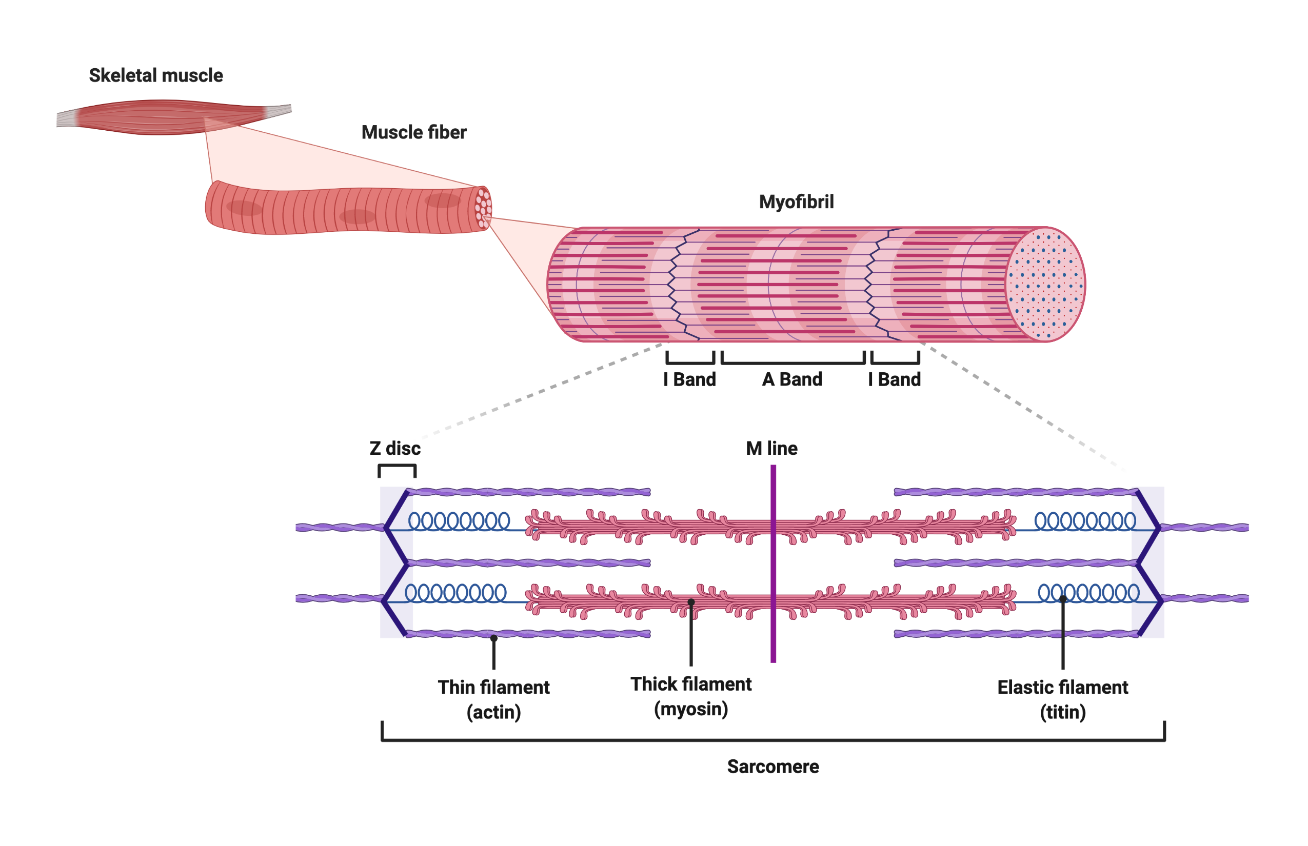
Muscle Fibers
In the hierarchical organization depicted in Figure 3.1 muscle fibers include a set of myofibrils arranged in parallel. The boundary of a muscle fiber includes the endomysium and sarcolemma (cell membrane of the muscle fiber). Myofibrils are surrounded by cellular components with critical roles to play when creating tension. Components such as extensions of the sarcolemma terminating at sacroplasmic reticula are critical for excitation and regulation; the mitochrondria are critical for energetics; and nuclei and associated protein manufacturing components such as the Golgi apparatus and ribosomes are critical for muscle maintenance and adaptation. For now we are focused on the muscle fiber components that generate tension. For the process of generating tension, the myofibrils are the functional building block of muscle fibers, and sarcomeres are the functional building blocks of a myofibril.
3.1.1 Anatomy of a Sarcomere
Figure 3.2 provides a view of the tensioning proteins of the sarcomere. The sarcomere includes all of the proteins between two Z-discs. These proteins are all actors that combine to create passive tension (resistance to positive length) and active tension through the cross-bridge cycle (sliding filament model).
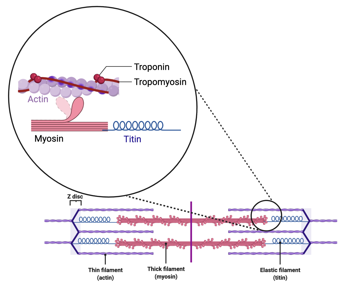
Primary and Secondary Roles of Primary Protein ”Actors”
-
•
Actin - primary role in active tension; secondary role in passive tension
-
•
Myosin - primary role in active tension; secondary role in passive tension
-
•
Troponin - Tropomyosin - primary role in active tension; secondary role in passive tension
-
•
Titin - primary role in passive tension; secondary role in active tension
Passive tension is created when the Z-discs are pulled apart. Active tension is created to pull the Z-discs closer together. Knowing the precise structure of each of these proteins is not necessary. Just keep in mind that their structure is of great importance. As with all proteins the structure is the foundation of function. Proteins form molecular machines that have very precise functions based on their very precise structure. Anything that alters the structure of a protein alters its function. Variations in pH and temperature that exceed certain boundary conditions, for example, have the capability of changing protein structure. Here we focus on the general structure of the proteins that allow the proteins to play their role in creating tension. The arrangement of these proteins with one another within the sarcomere is important. Everything you need to know about the structure and arrangement is based on the roles they play in the upcoming sections.‘
3.1.2 Structure and Arrangement of the Sarcomere During Changes in Length
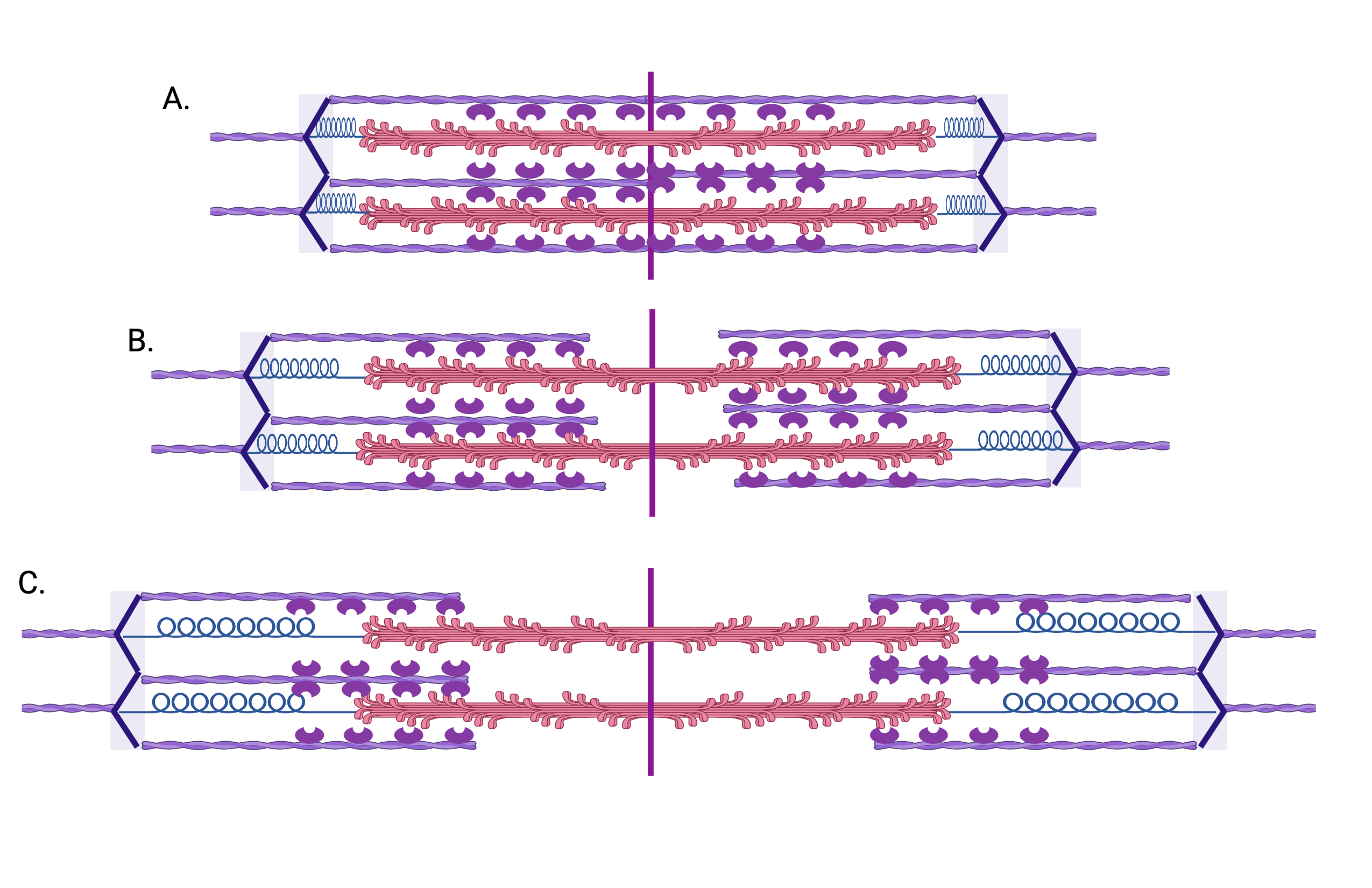
The structural change to the sarcomere at different lengths is the distance between the Z-discs (Figure 3.3). Increasing the distance between Z-discs reduces the space between the actin and myosin and, once at a certain length, starts to pull on the elastic structure of titin (unwinding of titin). Decreasing the distance between Z-discs results in increased space between actin and myosin, and recoil of titin to a point. Further decreases in distance between Z-disks results in crowding of potential actin myosin crossbridges and compression of titin. The implications of these structural changes are discussed in the sections on Active and Passive Tension.
3.2 Passive Tension
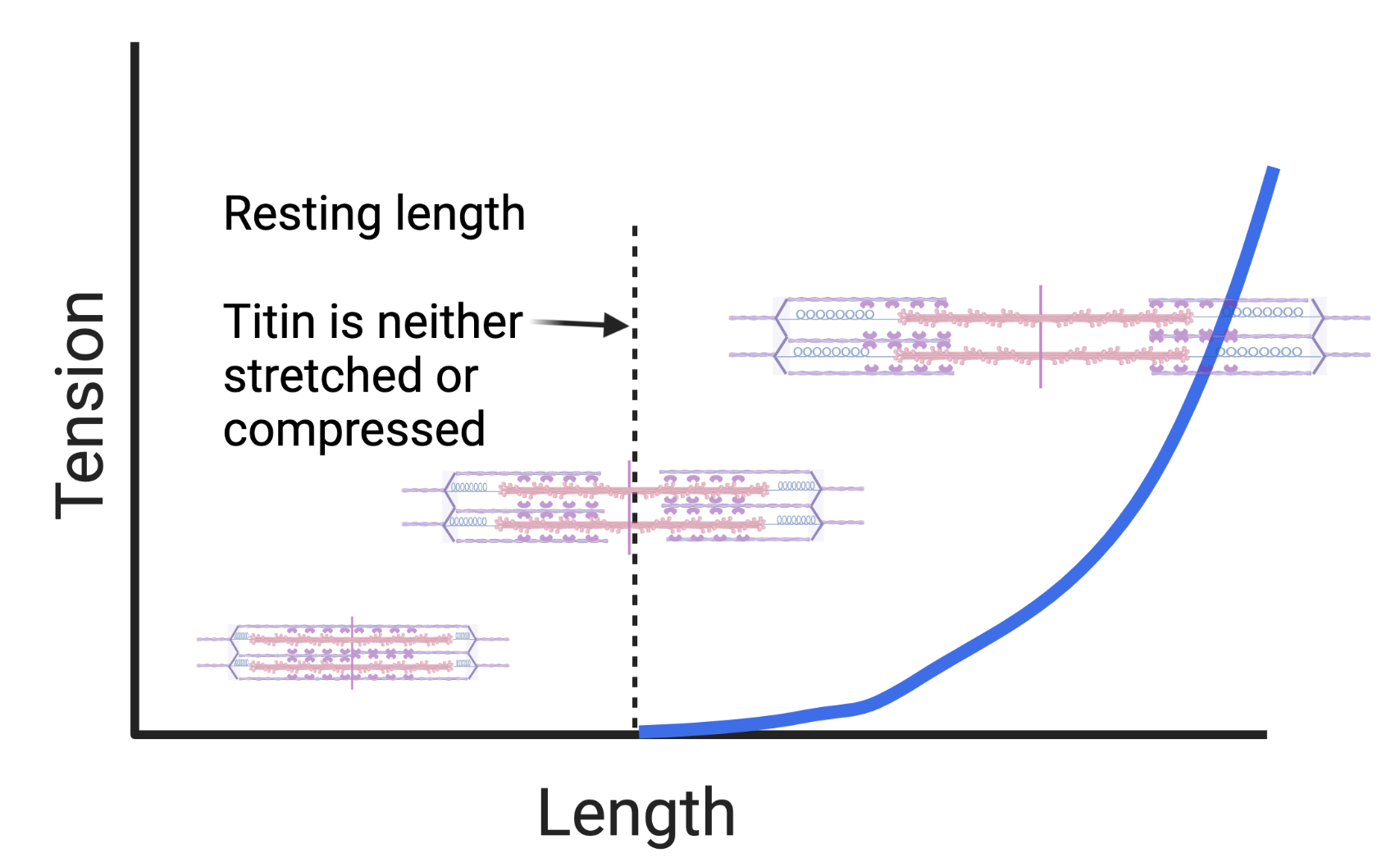
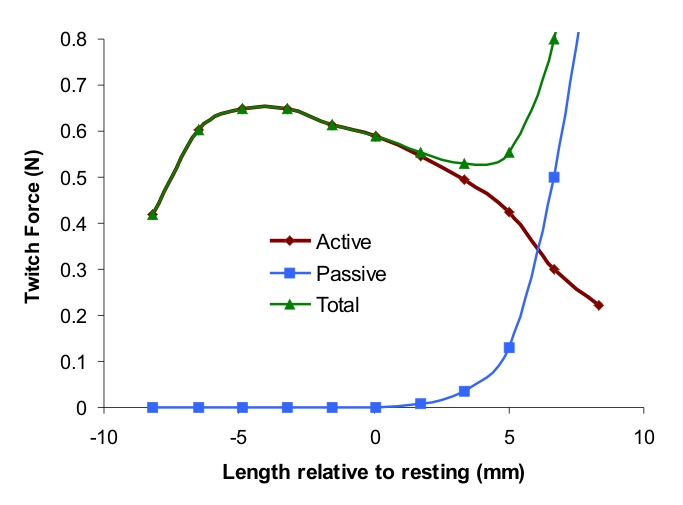
Passive tension is created when the Z-discs are pulled apart from one another. During positive length Z-discs of sarcomeres are pulled apart and muscle fibers increase in length. The amount of passive tension developed (force that resists lengthening) is a function of the relative length of the muscle. This is due to the relative length of titin and is the predominant component of the passive component of the length - tension relationship (Figure 3.4).
Muscle Tone
In an “inactivated” muscle there are some attachments between actin and myosin. The number of actin and myosin attachments influences the passive tension developed at various lengths. This is referred to as muscle tone. Muscle tone is the amount of tension in a muscle when it is not activated. Muscle tone is dependent on the length of the sarcomere since that length influences the number of possible actin myosin attachments. The presence of these actin myosin attachments in an inactivated muscle is primarily related to the underlying reasons for the attachments and is covered in later chapters.
3.2.1 Passive Tension Role In Movement
Passive tension can have a fundamental role in movement. Alterations in passive tension can therefore have a fundamental role in altered movement (kinesiopathology). When passive tension is developed the force by the muscle undergoing passive tension creates a torque across the joint. For example, when the hamstrings are lengthened and generate passive tension, that passive tension creates a flexion torque at the knee. If the hamstrings are then activated (start to generate active tension) then the total tension created is the sum of the active and the passive tension, which together create the force that creates the flexion torque at the knee. Muscles involved in gait (walking) can cycle through periods of passive tension and then use that passive tension to supplement active tension. For example, during running the calf muscles are lengthened while the foot is on the ground which increases passive tension. When it is time for that foot to propel the body the torque produced by the calf muscles is a combination of passive and active tension. In this situation, additional passive tension reduces the need for active tension and thus increases muscle efficiency (less energy is required, and therefore the muscle can sustain tension for a longer period of time).
3.3 Active Tension
To create active tension the muscle fiber transforms the chemical energy stored in adenosine tri-phosphate (ATP) into mechanical energy allowing myosin to actively slide actin from one actin-myosin crossbridge to another. This process is activated at the level of the muscle fiber and all of the events for energy transformation occur in the sarcomere. The result is tension that pulls (or attempts to pull) the Z-discs of the sarcomere closer together.
3.3.1 Sliding Filament Model
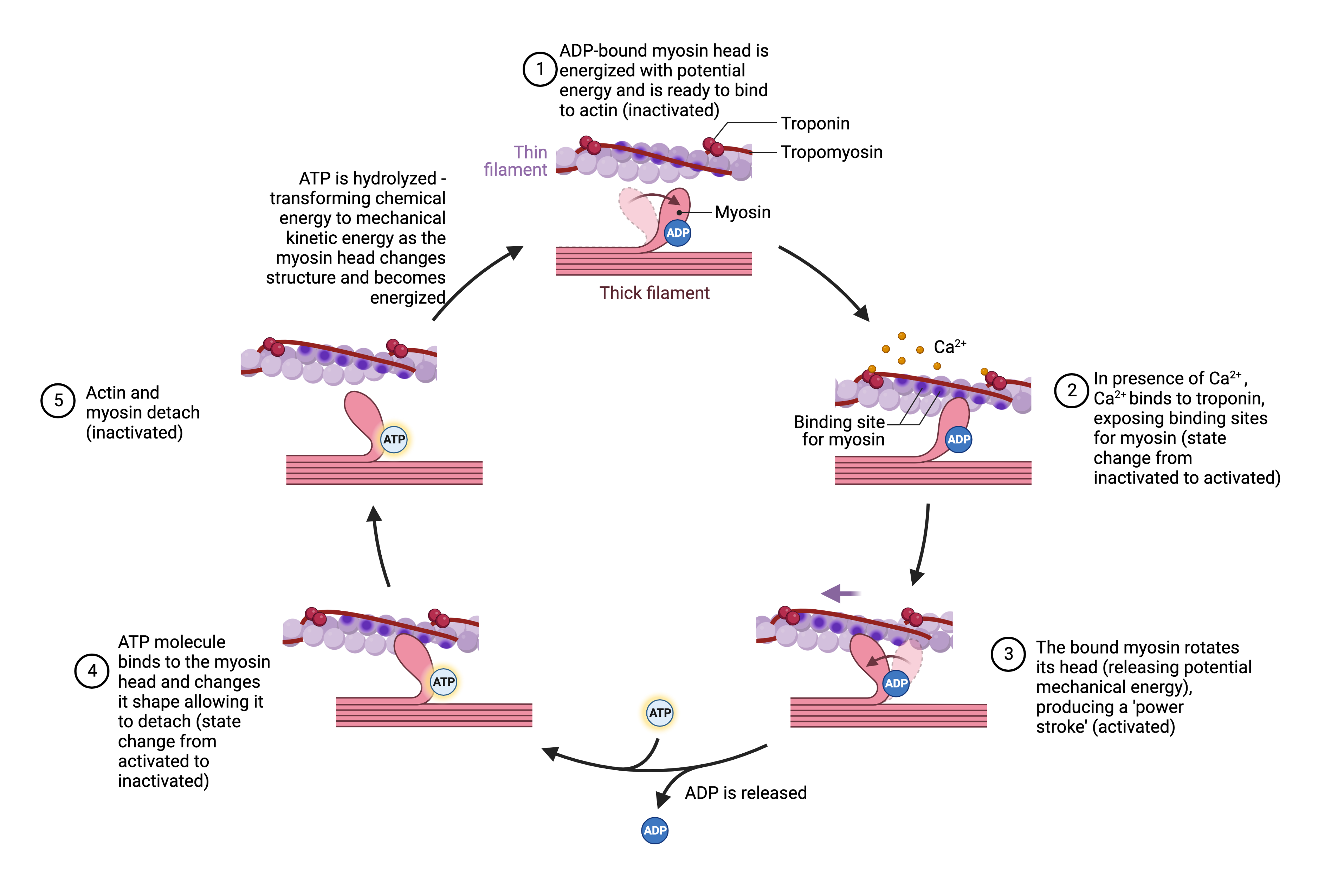
The sliding filament model of active tension is fairly well understood. There remains active investigation into the sliding filament process, the role of titin and the details of crossbridge kinetics. Figure 3.5 depicts the steps in the process of creating active tension. The steps are also enumerated below.
-
1.
ADP-bound myosin head is energized with potential energy (from the release of a phosphate from ATP) and is ready to bind to actin. While the myosin head is energized, it is inactivated.
-
2.
In the presence of calcium, calcium binds to troponin, moving tropomyosin and exposing actin binding sites so that myosin can bind to actin. In crossbridge kinetics terminology this is the change in state of the crossbridge from inactivated to activated.
-
3.
The bound myosin rotates its head (releasing potential mechanical energy), producing a ’power stroke’. The crossbridge remains activated.
-
4.
ATP molecule binds to the myosin head and changes its shape allowing it to detach from actin (state change from activated to inactivated).
-
5.
Actin and myosin are detached and not energized (inactivated)
Crossbridge Kinetics
The current understanding of the molecular basis for muscle contraction comes from Huxley’s (1957) kinetic model of the cyclic interaction between actin myosin crossbridges [huxley_muscle_1957]. Active tension behavior of a sarcomere (such as the force - velocity relationship described below) does not originate from the behavior of individual crossbridges, but from the collective action of all activated crossbridges in the sarcomere as they asynchronously go through the energy (ATP) dependent cycle described in the sliding filament model. In the crossbridge kinetics model the sarcomere has, at any given length, a number of possible crossbridges. The number of possible crossbridges is the crossbridge interaction space. Each possible crossbridge has two states, activated and inactivated. The active tension of a sarcomere is directly proportional to the number of crossbridges in the activated state. The number of activated crossbridges is influenced by both the concentration of calcium in the sarcomere and the length of the sarcomere. The sliding filament model describes one cycle of crossbridge kinetics (one cycle of inactivation to activation to inactivation).
Introduction to Twitch
A twitch is the fundamental unit of active tension. It is a spike in active tension generated when crossbridges are activated from one excitation of a muscle fiber. A twitch can be considered at the level of the sarcomere, myofibril, fiber and muscle. A twitch in situ is perceptible but not functional. If many muscle fibers twitch at the same time it may be experienced as a spasm, though not long lasting. Sustained spasms (myotonia) involve more than a twitch. A twitch can be thought to equate to one cycle of crossbridge kinetics. The force of a twitch is directly proportional to the number of crossbridges in the activated state, meaning if there are more crossbridges cycling into the activated state there is more tension. Experimentally, all sarcomere twitches will tend to produce the same tension when performed at the same length and when separated by enough time. Myofibril twitches can have small variations in tension depending on how many of its sarcomeres have crossbridge cycling from the stimulus provided. Muscle fiber twitches can vary greatly in the amount of tension during a twitch because there is greater potential for variation in the number of sarcomeres involved in crossbridge cycling. Experimentally the number of sarcomeres involved in cross bridge cycling depends on the amount of excitation stimulus provided.
Introduction to Tetany
When a sarcomere is repeatedly excited the resultant twitches from each excitation start to fuse and the spikes in tension from each twitch get smooth while the overall tension increases. This transition to a smooth rise and even maintenance of tension from a sarcomere is tetany. The greater the number of excitations the greater the tension developed in tetany because the result is a greater number of crossbridges in the activated state at any given moment. The amount of tension developed reflects the ability to attain tension, and the period of time tetany can last during repeated excitations is the ability to sustain tension. In Chapter 4 the concepts of twitch and tetany are the bridge between muscle excitation and crossbridge kinetics. In Chapter 5they are used to connect muscle regulation to excitation.
3.3.2 Active Component of the Length Tension Relationship
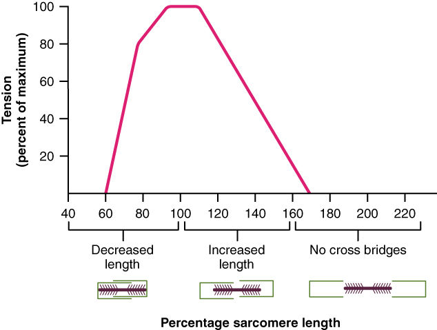
Since the sarcomere crossbridge interaction space varies with its length 111Distance between the Z-discs the number of activated crossbridges also varies with its length. Therefore, under conditions of maximal activation (high calcium concentrations), the active tension varies with sarcomere length. The crossbridge interaction space decreases as the distance between the Z-discs increases or decreases from an ideal distance. The optimal distance between the Z-discs (optimal sarcomere length) is defined as the length that maximizes the crossbridge interaction space.
The active component of the length tension relationship was originally measured using isolated muscle fibers under isometric conditions at different lengths. However, we tend to consider the impact of the underlying phenomenon during activation that includes dynamic changes in length. As the distance between the Z-discs decreases, such as during negative length (concentric) or when muscle activation begins while in a shortened length, the actin-myosin interaction space is decreased because of the actin binding sites increase in concentration with a limited number of myosin heads (near the M-line) which reduces the number of potential crossbridge sites. This reduces the active tension. As the distance between the Z-discs increases, such as during positive length (eccentric) or when muscle activation begins while in a lengthened length, the actin-myosin interaction space is decreased. This occurs because parts of actin are not in proximity with myosin which reduces the number of crossbridge sites. This result is a reduction in active tension. The active component of the length tension relationship is depicted in Figure 3.6.222The depiction of the actin myosin crossbridges in this particular rendering includes overlap of the actin during the shortened range. Whether there is such overlap vs. crowding of sites near the M-line is still an open discussion, or at least as far as the author is aware.
3.3.3 Length-Tension Relationship (Active & Passive Components)
The full length tension relationship of a sarcomere, and a myofibril, is depicted in Figure 3.7. This relationship is based on experiments with myofibrils removed from their anatomical locations and maximally activated under isometric (zero length) conditions. The length tension relationship of any particular muscle in situ may vary from this general relationship based on factors such as the normal resting position of the muscle with anatomical attachments, the pennation arrangement, whether the conditions of maximal activation are met (rarely), whether the conditions of zero velocity is met (rarely during functional movement), or whether the muscle crosses more than one joint. Two situatinos deserve special mention.

Manual Muscle Testing
During manual muscle testing (MMT) physical therapists are taught about the ideal joint range of motion (ROM) for testing. These ROMs are typically positions that put the muscle at a length intended to maximize tension based on the active component of the length tension relationship.
Active & Passive Insufficiency & Sufficiency
A special situation arises in muscles that have attachments crossing more than one joint. The ROM of both joints influences the muscle length. Since muscle length influences both active and passive tension the position or movement of both joints influences active and passive tension. Active insufficiency occurs when the active tension a muscle creates is impacted by the position or movement of the joints (at least 2) that is crosses. For example, if you attempt to extend the hip and flex the knee at the same time using active tension your ability to generate active tension declines quickly due to the fact that the hamstring sarcomere Z-discs are getting closer and actin-myosin interaction space is decreasing.
Passive insufficiency occurs when the passive tension a muscle creates is impacted by the position or movement of the joints (at least 2) that it crosses. For example, if you attempt to flex the hip and extend the knee at the time time, passive tension develops due to the hamstring sarcomere Z-discs getting further apart and titin is resisting further changes to its length.333Please note that the focus here on titin is for simplicity. Other connective tissues within the muscle contribute to this passive tension. It is also possible that extra-muscular connective tissue such as fascia can contribute to the passive tension developed during passive insufficiency since these movements tend to not serve critical movement functions. They tend to be movements contrary to what would be considered critical movement functions. Active and passive sufficiency is the principle that most functional movements avoid active and passive insufficiency. Most functional movements tend to use multi-joint muscles in a way that optimize their tension generating capabilities based on the length tension relationship. For example, during squatting and climbing, the hamstring sarcomere length does not change much since these movements couple hip flexion with knee flexion (one direction of movement), and hip extension with knee extension (the other direction of movement).
3.3.4 Necessary Molecules
Most of the molecules involved in the sliding filament model are proteins in the sarcomere. There are two molecules that make important appearances and deserve special mention as a preface to where we are heading as we progress toward connecting muscle physiology to clinical physiology.
Calcium
Calcium() enters the scene to get things started. It triggers events that lead to the crossbridge state change from inactivated to activated. It is the signaling molecule utilized to activate the process of the sliding filaments for active tension. It is stored in an organelle in close proximity to the sarcolemma called the sarcoplasmic reticulum. enters the cell once the sarcolemma and sarcoplasmic reticulum is excited and is an important part of Chapter 4 on Excitation. is the connecting molecule in the process called “excitation - contraction coupling” which we call “excitation - activation coupling”. Having sufficient for the muscle fiber is so essential that the endocrine system helps ensure it is always available, even at the expense of in the bones.
Adenosine Triphosphate (ATP)
Adenosine Triphosphate (ATP) provides the chemical energy that is transformed into mechanical energy by the molecular motors within the sarcomere. ATP is kept in limited quantities inside the muscle cell. It is used to energize cellular processes that require energy (do not confuse cellular processes with chemical reactions). Examples of cellular process include transcription and translation or a pump that moves ions across a membrane to form a gradient. Chemical reactions are transformations that proceed bidirectionally with one direction usually favored by mass balance and enzyme catalysts. Since ATP is stored in limited quantities (enough for seconds of myosin energizing), the muscle fiber must be able to convert other forms of energy (usually chemical energy) into ATP. That is the primary topic of Chapter 6 on Muscle Energetics.
3.4 Force & Velocity
There is a relationship between force and velocity. The relationship between them is considered from two perspectives: the Force - Velocity (FV) and the Velocity - Force (VF) Relationships. This chapter introduces the concept and these two perspectives (FV & VF), and considers the how sarcomeres contribute to this understanding. By force we are referring to force being measured that is the result of muscle tension (total tension). By velocity we are referring to changes in length over time.
length and velocity are related based on Equation 3.1:
| (3.1) |
Velocity involves changes in length which makes the force velocity relationships different from length-tension relationships. Length tension relationships are experimentally observed under conditions of isometric (zero length) conditions and is theoretically extended to dynamic (non zero length) conditions. Force velocity relationships must be experimentally observed under dynamic (non zero length) conditions. Since velocity involves changes in length, the force velocity relationships must include considerations related to the implications of the length tension relationship. Even though the length tension relationship is observed under conditions of isometric activation, what the length tension relationship represents (changes in sarcomere structure that result in changes in passive elastic and active tension) is occurring during a change in sarcomere length.
Movement Energetic Perspective
Before proceeding with the FV and VF relationships it is important to point out that these relationships can be differentiated from a third perspective - the movement energetic perspective. Movements are the sum of torques acting across joints with a larger imbalance between torques producing a “faster” movement, and keeping in mind that torques are generated by force which is generated by active muscle tension, and that active muscle tension is proportional to the number of activated crossbridges, and that the number of activated crossbridges is proportional to the amount of ATP being utilized, we can conclude that “faster” movements utilize more ATP. Therefore they are more energetic. The movement energetic perspective is discussed in Chapter 6 on Muscle Energetics. In summary, it is why running at a faster pace makes you breath heavier than a slower pace; and why certain running paces (sprinting) are not possible for very long. 444The movement energetic perspective is not traditionally considered as part of the FV and VF relationships.
3.4.1 Force - Velocity & Velocity - Force Relationships
Velocity - Force Relationship
We consider the VF Relationship under the condition of no resistance. In the VF relationship force is a function of velocity. The muscle is not resisted 555By anything of any significance beyond the resistance of joint friction, limb mass and air resistance. The point is that, in this situation, velocity is not impacted by the necessity for the muscle to generate enough active tension to overcome the force of the resistance. and we consider the force that can be achieved with varying levels of velocity. The independent (manipulated) variable is on the X axis, and as the dependent (observed) variable is on the Y axis. We go into more depth for the VF relationship in Chapter 5 on Muscle Regulation since the phenomenon involves variations in muscle fiber types (heterogeneity between muscle fibers).
Force - Velocity Relationship
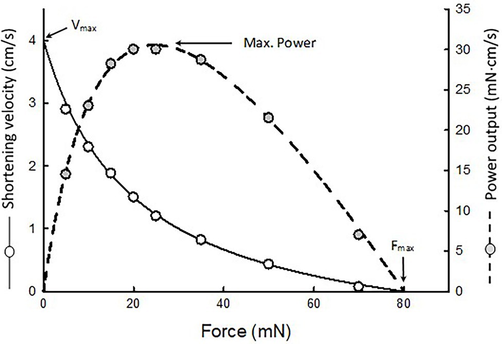
We consider the FV Relationship under the condition of resistance. In the FV relationship velocity is a function of force (as in the force required to move a load). The relationship between force and shortening velocity is visualized by plotting the velocity of a shortening muscle as a function of the load (or force) pulling on the muscle [seow_molecular_2022]. The muscle is resisted and we consider the velocity that can be achieved with varying levels of resisted muscle force. The independent (manipulated) variable is on the X axis, and the dependent (observed) variable velocity is on the Y axis. FV has an inversely proportional hyperbolic relationship and was first described by Hill [seow_hills_2013]. Because power is the product of force and velocity it has a parabolic relationship which achieves its peak at neither the extreme of velocity or force [seow_hills_2013].
Force Velocity Relationship during Shortening (Concentric)
Figure 3.8 shows the FV relationship during concentric (shortening) activation with power. It is a consistent finding that peak power occurs between 20 and 30% of peak force.
There are two factors contributing to the FV relationship during concentric activation. First, is the fact that a concentric activation requires the muscle generate more torque than the resistance (load). When someone generates just enough torque, they generate a minimal (non zero) velocity. As load is reduced, the maximal active tension of the muscle creates an abundance of torque and this results in greater velocity.
Second, under the condition of attempting to move as fast as possible, as the load on and the force generated by a muscle increases, there is an increase in the velocity. The drop in force is due to the drop in load, which then allows for a higher velocity. However, it is also true that at the higher velocity there is a lower capability for generating force (the VF relationship). This is due to two factors that can be traced back to crossbridge kinetics. First, with increased velocity of shortening the crossbridge interaction space decreases rapidly which brings the sarcomeres into a shorter length and reduces the tension they can create (i.e. length tension relationship). Second, with increased velocity of changes in length there is less time for crossbridges to be in their activated state, which means at any moment there is a lower number of possible crossbridges in the activated state.
Experience of the FV relationship during concentric activation
The FV relationship during concentric activation is something people can easily relate to and have experienced. There is a minimal velocity associated with any activity people perform with maximal resistance (force). Note that minimal velocity varies between activities. For example, the minimal velocity of a dead lift is lower than the minimal velocity of a power clean.
Force Velocity Relationship during Lengthening (Eccentric)
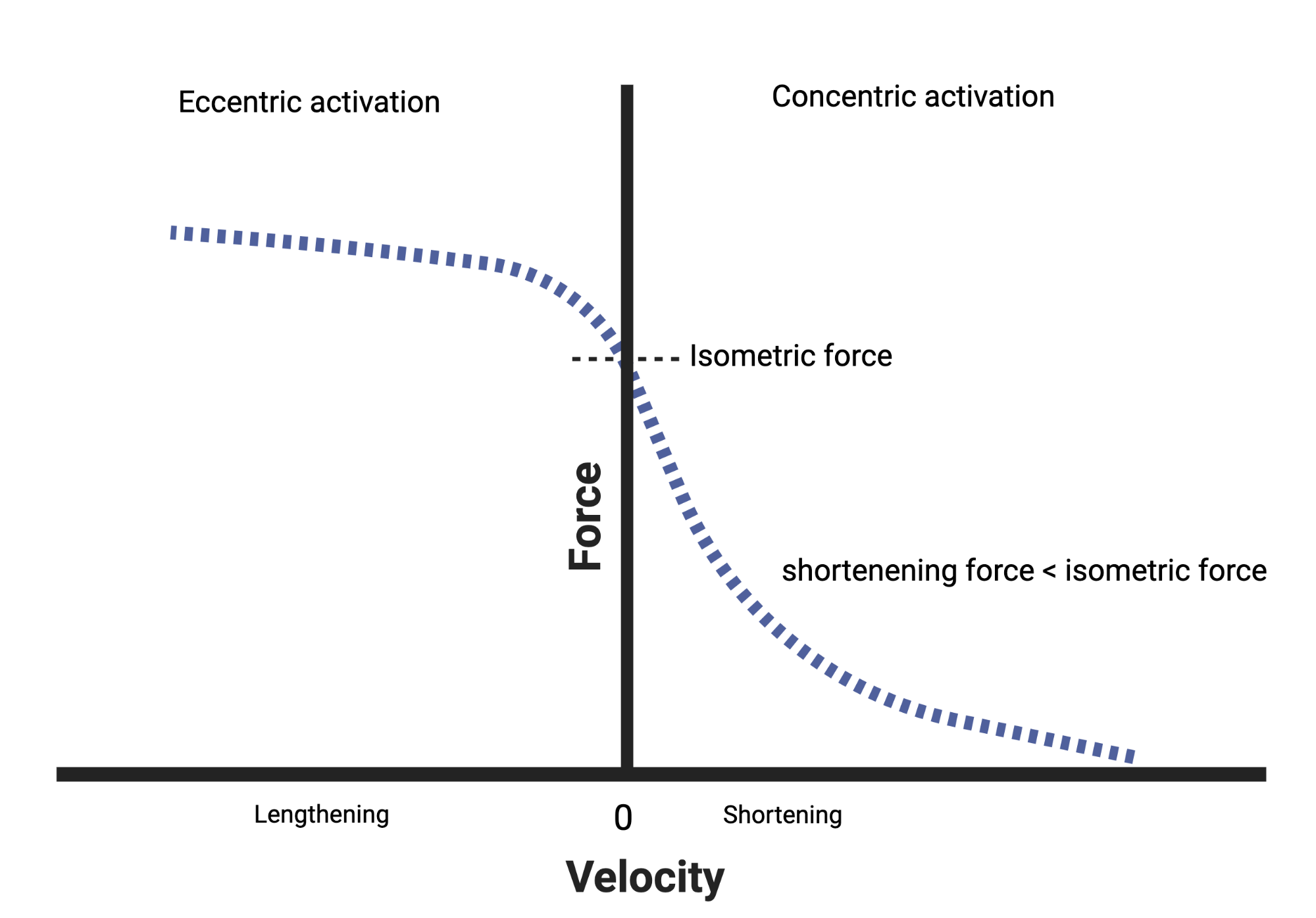
The FV relationship for eccentric (lengthening) activation is depicted by plotting velocity on the X-axis and force on the Y-axis since it requires a change in the direction (sign) of velocity that occurs as you go from concentric to eccentric activation. We will see this same axis flip when we consider the VF relationship in Chapter 5.
Figure 3.9 shows the relationship between force and velocity during both shortening (right side of Y axis, positive velocity) and lengthening (left side of the Y axis, negative velocity). There is a rapid increase in force at the initial transition point. The force (load, resistance) is now overwhelming, that is exceeding, the capacity of the sarcomere to generate force. But it is just exceeding which engages the passive elements of the sarcomere (titin) to contribute to force and has not yet resulted in rapid loss of the crossbridge interaction space or time for crossbridge activation. The force that can be generated by the muscle (in opposition to the external force (load, resistance)) quickly plateaus once titin has been fully engaged and the increased velocity diminishes time for crossbridge activation and the lengthening of the muscle results in less crossbridge interaction space (again, due to the length tension relationship).
Experience of the FV relationship during eccentric activation
The FV relationship during eccentric activation is something people can easily relate to and have experienced. Very simply, as the load someone attempts to hold (but cannot actually hold) increases, the velocity of movement also increases.
3.5 Summary
In this chapter we have covered the micro anatomy of muscle fibers at sufficient depth to understand the mechanisms behind active and passive tension as well as several concepts relevant for physical therapy practice such as the length tension and force velocity relationships. The sarcomere is the functional unit of the muscle fiber for active tension. Within the muscle fiber, the parts and mechanisms within a sarcomere can generate active tension by converting chemical energy into mechanical energy, or passive tension by resisting change to length. Active tension involves a sequence of activation events repeated over and over. The sequence of activation begins with excitation. Therefore, our next step is to consider excitation.
[heading=subbibintoc]
Sample Questions
-
1.
Which structures do not have a primary role in active tension?
-
2.
Which structures do have a primary role in active tension?
-
3.
What is the correct sequence for generating active tension?
-
4.
Maximal quadriceps shortening velocity during knee extension at an 80% maximal tension load would be a (higher or lower) velocity than during knee extension at 90% maximal tension load.
-
5.
Maximal quadriceps tension during knee extension at 360 degrees per second would be (higher or lower) than during knee extension at 30 degrees per second.