Chapter 8 Renal Clearance
Updated on September 4, 2026
This chapter covers the concept of mass balance with a particular focus on water and electrolytes. The mass balance of water and electrolytes are supported by renal clearance which includes filtration, reabsorption and secretion for the formation and excretion of urine. Water and electrolyte mass balance are important processes in the support of plasma (and therefore blood) volume; as well as the content and concentration of electrolytes in the extra-cellular fluid. Alterations in the plasma volume and concentration influence the interstitial fluid and the ICF. The regulation of body fluid volume and composition is accomplished by acting directly on the plasma. The kidneys, and systems such as the gastrointestinal, metabolic, and pulmonary, participate in mass balance and the regulation of the body fluid volume and composition.
Objectives include:
- 1.
Explain mass balance.
- 2.
Relate mass balance to the ICF model and physical therapy practice.
- 3.
Summarize the role of the kidneys.
- 4.
Explain the role of the kidneys in mass balance.
- 5.
Explain the functional anatomy of the kidney and nephron and the unique arteriole system including possible symptoms and signs of kidney conditions that should be part of the differential diagnosis of musculoskeletal back pain.
- 6.
Explain renal clearance.
- 7.
Explain urine formation and excretion by micturition and common causes of incontinence
- 8.
Explain glomerular filtration, factors that influence GFR and the use of BUN and creatinine to assess GFR.
- 9.
Explain the role of tubular reabsorption and secretion and how they influence the excretion rate.
- 10.
Describe renal regulation of sodium, potassium, potassium, calcium, acid base balance.
- 11.
Explain renal regulation, and integration of blood volume and osmolarity including the role of renin, angiotensin II, aldosterone and ADH.
- 12.
Explain the response to positive and negative water balance.
- 13.
Explain disturbances of fluid volume & osmolarity.
- 14.
Explain rhabdomyolysis and the possible role of water homeostasis on frailty in aging.
8.1 Mass Balance
Mass balance applies the conservation of mass and energy to the analysis of physical systems. It considers the mass and energy entering (inputs) and leaving a system (outputs). Physical therapists help individuals maximize activity and participation such as activities of daily living (ADLs). ADLs are largely based on the need for water and nutrient input, and hygienic output of waste. To use the terms from the ICF model introduced in Chapter 1, the inputs of food, drink and air require activity and participation (function). Many activities of daily living (ADLs) are based on the functional activities that provide appropriate inputs and hygienic outputs required for physiological mass balance.
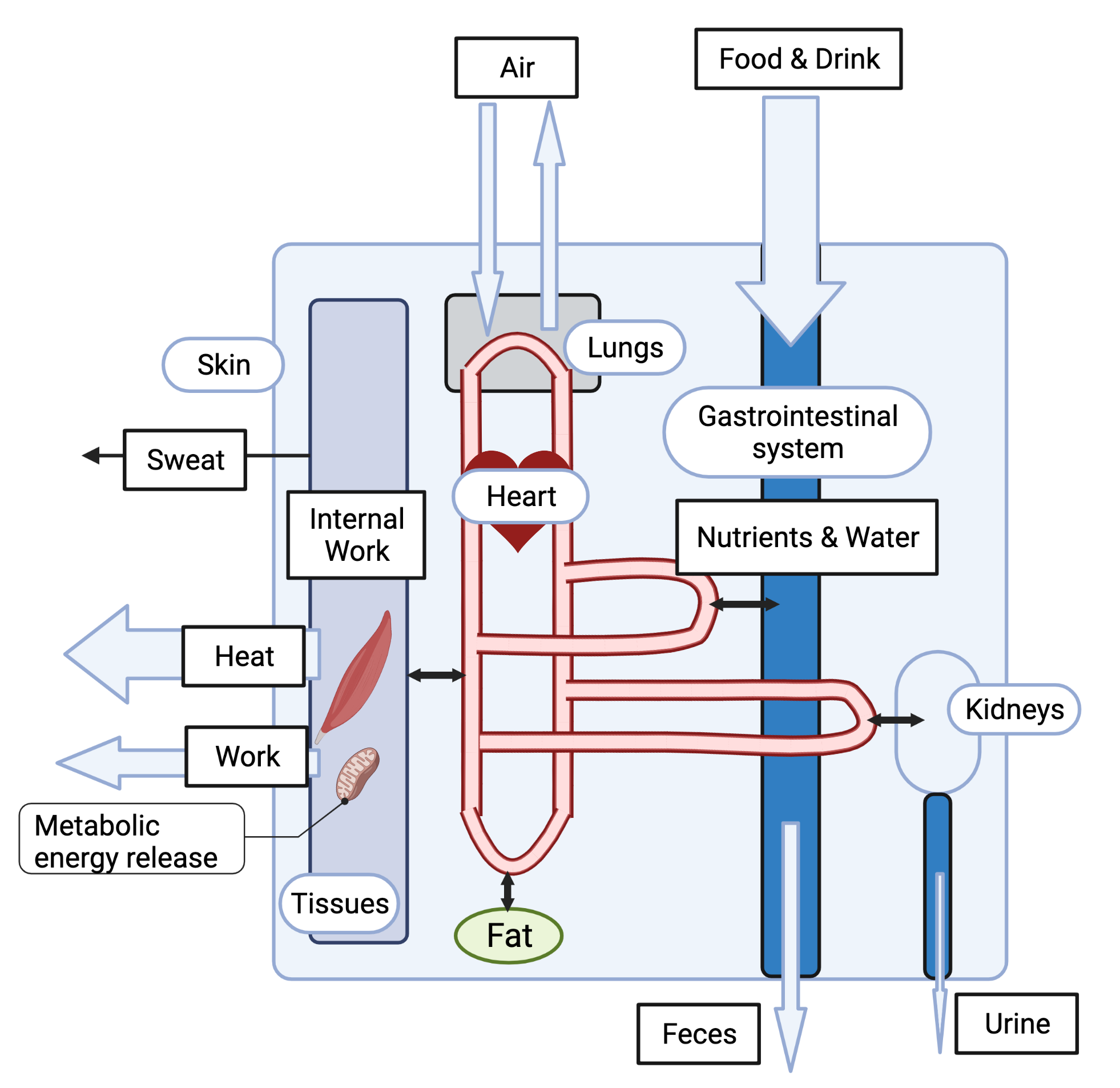
The inputs of food, drink and air ultimately provide the water, nutrients (macronutrients and micronutrients) and oxygen that are utilized for physiological processes. An overview of physiological mass balance is provided in Figure 8.1. How these are used, whether they can be recycled, and the ways in which they are lost influences the time scale of the need for inputs. When describing outputs the term incidental loss refers to loss that occurs without purpose. Incidental loss is contrasted with intentional loss. Intentional loss is loss for a purpose, such as the loss of water in urine to regulate water balance. An example of incidental loss is the loss of in the gastrointestinal tract due to sloughing of cells from the mucosal wall; and the loss of water with breathing. It is interesting to consider whether losses such as in sweat is incidental or intentional. It is clear that water loss in sweat is intentional - for the purpose of thermoregulation. While loss is not needed for thermoregulation, its loss may help sustain ECF osmolarity. There are several additional examples that go beyond what we can consider here.
8.1.1 Oxygen Mass Balance
Oxygen is used regularly and relatively quickly with very little storage (myoglobin) and no recycling, making the input of air with a regular requirement. Life is not sustainable for prolonged periods without the input of air with over the time scale of a few minutes in most individuals. At nearly the same rate, and at times at a slightly higher rate than oxygen utilization carbon dioxide is produced as a waste product of mitochondrial respiration. Input and output of air allows for both the input of oxygen, and the output of carbon dioxide.
8.1.2 Nutrient Mass Balance
When discussing mass balance of nutrients, which includes the consideration of diet and nutrition since it is about the input of nutrients, it is common to break nutrients into two main classifications. Macronutrients are larger molecules that can be utilized for energy purposes. They have an energetic (caloric) value in addition to other physiological roles they fulfill. Macronutrients include carbohydrate, fat and protein. Micronutrients are small molecules and in some cases simply elements or minerals. They do not have an energetic value but are essential to many physiological processes. For muscle physiology it has already become clear that , and are essential micronutrients.
Background: Semantics of Stuff
The same stuff - such as glucose, calcium, sodium and potassium - can be referred to using different words based on context. Glucose is a macronutrient, but once ingested and absorbed and present in the blood or cells it tends to be referred to as substrate. Micronutrients are dietary inputs. They can referred to as electrolytes when considering the role they have in determining the osmolarity of a solution (e.g., ). They are considered ions considering the role they have in membrane potentials (e.g., ). They are considered signaling molecules (or just molecules) when considering the role they play in activation (e.g., ).
Macronutrients are routinely utilized for energy and structural health of cells, tissues and molecules such as hormones and neurotransmitters. They are also stored purposely. Fat is stored most abundantly as adipose tissue. Carbohydrate as glycogen. Protein is available from the breakdown of any existing proteins when in need of this substrate. Proteins can be recycled. If a protein is broken down into amino acids and used to build an enzyme that is needed; it can be broken down again and used to build another enzyme. Fat and carbohydrate utilized in structures can be recycled. Macronutrients utilized for energy lose their structure and cannot be recycled, they must be replaced. The digestion of food to extract and absorb usable substrates ultimately yields waste that remains in the gastrointestinal (GI) tract (for example, insoluble fiber) and is removed as feces.
The lining of the GI tract not only absorbs all nutrients (macro and micro), but there is some incidental loss of micro-nutrients (electrolytes and minerals) such as in the feces from the GI secretions and normal wear and tear of the cells and tissue that line the GI tract. A small amount of incidental loss of micronutrients such as and occurs in the sweat and urine. Since the input of these micronutrients can easily exceed the incidental loss, there can be intentional (regulated) loss through the renal filtration and secretion processes to maintain , and homeostasis.
8.1.3 Water Mass Balance
Water is the source of the fluid environment that the biochemical processes of life occur, and transport of essential nutrients flow through the circulation and micro-circulation. Without water the blood plasma, interstitial fluid and intra-cellular fluid would not be fluid. The fact that they are fluid enables nutrient movement. The amount of water in the body also influences the concentration (and therefore osmolarity) of these fluid spaces. The amount of water influences the blood volume. If blood volume is too high, blood pressure increases and edema may result. If blood volume is too low due to a loss of water, viscosity increases which increases resistance to circulation. Problems occur when there is too little water (dehydration) or too much water (elevated blood volume, edema). The water in the body is always in use, being filtered between compartments in support of the movement of nutrients and electrolytes as well as by-products (waste). In this way it is recycled - there is no need for a complete overturn of water in the course of a day. However, the amount of water that must be taken in (input) should equal the amount of water that is lost in a day (output). Water loss occurs through four routes, two incidental and two intentional. Incidental water loss occurs with ventilation of air out of the body; and a small amount of water is routinely lost in the feces. Evaporation of water from the skin is a mechanism of cooling the body and sweating is a route of loss. Intentional loss of water in sweat is highly variable based on the temperature and activity levels which influence the need for cooling. Renal filtration and excretion requires a loss of water in urine for the removal of metabolic waste. Of these four routes for the output of water sweating is highly variable and can be partially regulated through behavioral adaptations. Renal is also highly variable and is regulated through physiological processes. The maintenance of normal water volume, and as a result plasma volume, is buffered by fluctuations in renal output in response to water input. Because there is a continual regular loss of water (breathing and sweating), and a necessary regular loss of water for waste removal (feces and urine), there is a regular need (demand) for water input. While the kidneys can make more or less concentrated urine which includes less or more water (respectively), the body must make urine regularly to filter the blood and keep it free from the toxicity of metabolic waste products.
Blood Volume Composition
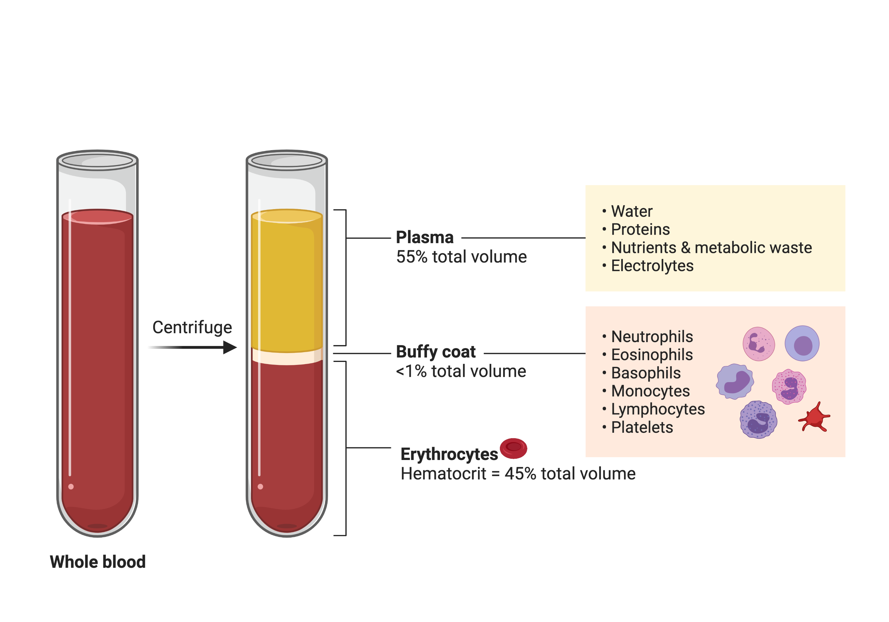
Blood volume includes plasma (approximately 55%) and cells (See Figure 8.2). Most of the plasma is water. Most of the cells are red blood cells (RBCs) and are also called erythrocytes. RBCs contain hemoglobin (HgB). RBCs are the oxygen carrying cells in blood. Hematocrit (HcT) reports on the percentage of blood volume that contains RBCs. Since other cells occur in relatively low volume, it is acceptable to consider plasma as simply 1 minus HcT when HcT is reported as a fraction (HcT of 45% is 0.45). If HcT is 0.45, then 1-0.45 = 0.55, which means 55% of the blood is plasma. The volume (size) of RBCs influence HcT. If there are the same number of RBCs but each has more volume then the HcT is increased (assuming plasma volume has not changed). If the RBCs have less volume then the HcT is decreased (assuming plasma volume has not changed).
8.2 Renal Function Overview
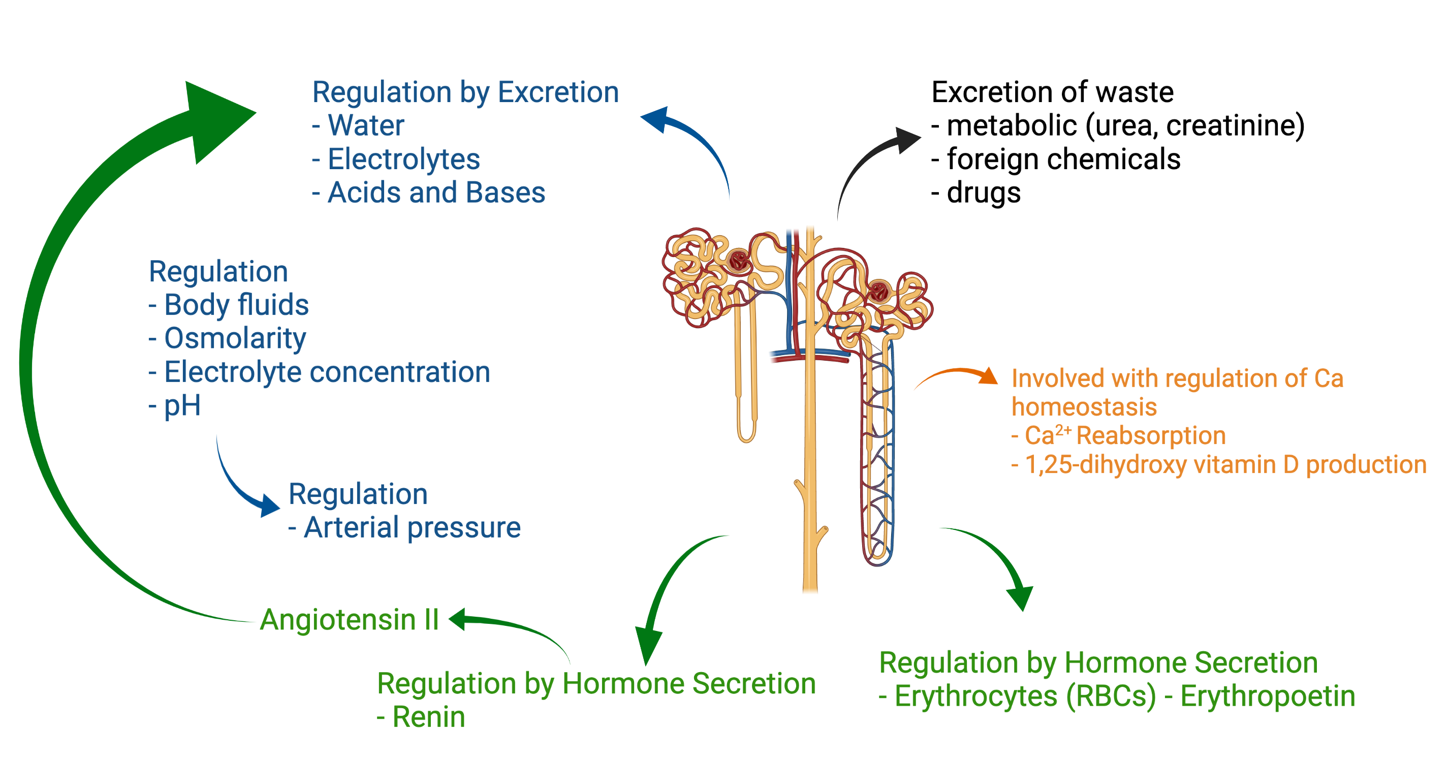
Renal function relies on the kidneys. The kidneys have several important physiological functions summarized below and in Figure 8.3.
-
1.
Excretion: The kidneys ensure that harmful substances are excreted in urine. Harmful substances can be absolutely harmful (waste products such as urea and creatinine), or relatively harmful (electrolytes when they exceed their normal ranges).
-
2.
Regulation & Buffer by Excretion: The kidneys regulate and therefore buffer the mass balance of plasma, and therefore body water and electrolyte amounts. Through both water and the electrolyte content they regulate electrolyte concentration.
-
3.
Regulation by Excretion: Through the regulation of plasma volume the kidneys provide long term regulation to blood pressure.
-
4.
Regulation by Hormone Secretion: The kidneys synthesize and secrete three hormones:
-
(a)
Renin for the regulation of blood pressure
-
(b)
Erythropoietin for the regulation of red blood cells
-
(c)
1,25-dihydroxycholecalciferol for the regulation of
-
(a)
Functions 1, 2 and 3 and the endocrine function of renin are covered in this chapter.
Functions 1 and 2 are accomplished through renal clearance. Renal clearance is a general concept that describes the rate that substances (including water) are removed from plasma. Renal clearance is accomplished as blood passes through the renal capillaries and undergoes filtration. Filtration in the renal capillaries greatly exceeds the filtration in systemic capillaries and therefore is also referred to as ultrafiltration. The fluid that is filtrated out of renal capillaries is referred to as ultrafiltrate. Ultrafiltrate proceeds through the renal tubule while the more selective processes of reabsorption and secretion fine tune its content until it is eventually urine and headed for clearance. In total, renal clearance ensures that harmful substances are excreted (filtered and not reabsorbed); that the correct volume of water and electrolytes remain in the plasma (filtered and then reabsorbed as necessary, or not filtered and secreted as necessary).
8.2.1 Functional Anatomy
The kidneys are in the retroperitoneal cavity of the body. Given their location and the high prevalence of back pain in physical therapy practice it is important to consider the possibility that back pain is related to the kidneys.
Background: Kidney Pain
Kidney pain is pain from disease or injury to a kidney. Kidney pain or discomfort may manifest as a dull, one-sided ache in the upper abdomen, side or back. Back pain that includes upper abdomen or side components and that does not respond to provocations that would typically provoke a musculoskeletal source of pain such as movements (active & passive tension) should be considered. The patient should be questioned regarding a history of the common causes of kidney diseases such as alcohol use, diabetes, high blood pressure, family history or a history of kidney stones. Fever and changes in urinary habits or urinary symptoms often accompany kidney pain and are therefore important considerations when ruling out a kidney as a source of back pain.
Each kidney is divided into two regions: the outer cortex and the inner medulla. Comprising these two region are tube-like structure called nephrons - the functional unit of the kidney. Each kidney can contain a million individual nephrons with each nephron serving a vital role in reabsorption, secretion, and filtration of solutes. At its simplest, each nephron contains a renal corpuscle (glomerulus surrounded by Bowman’s Capusle) and renal tubule. The glomerulus is a capillary network and the primary site for plasma filtration and the formation of urine. Distributed on both ends of the glomerulus are the afferent and efferent arterioles, which provide inflow of filtrates to the glomerulus and outflow of filtrates to the upper portion of the nephron - the Bowman’s Capule (or Bowman’s Space).
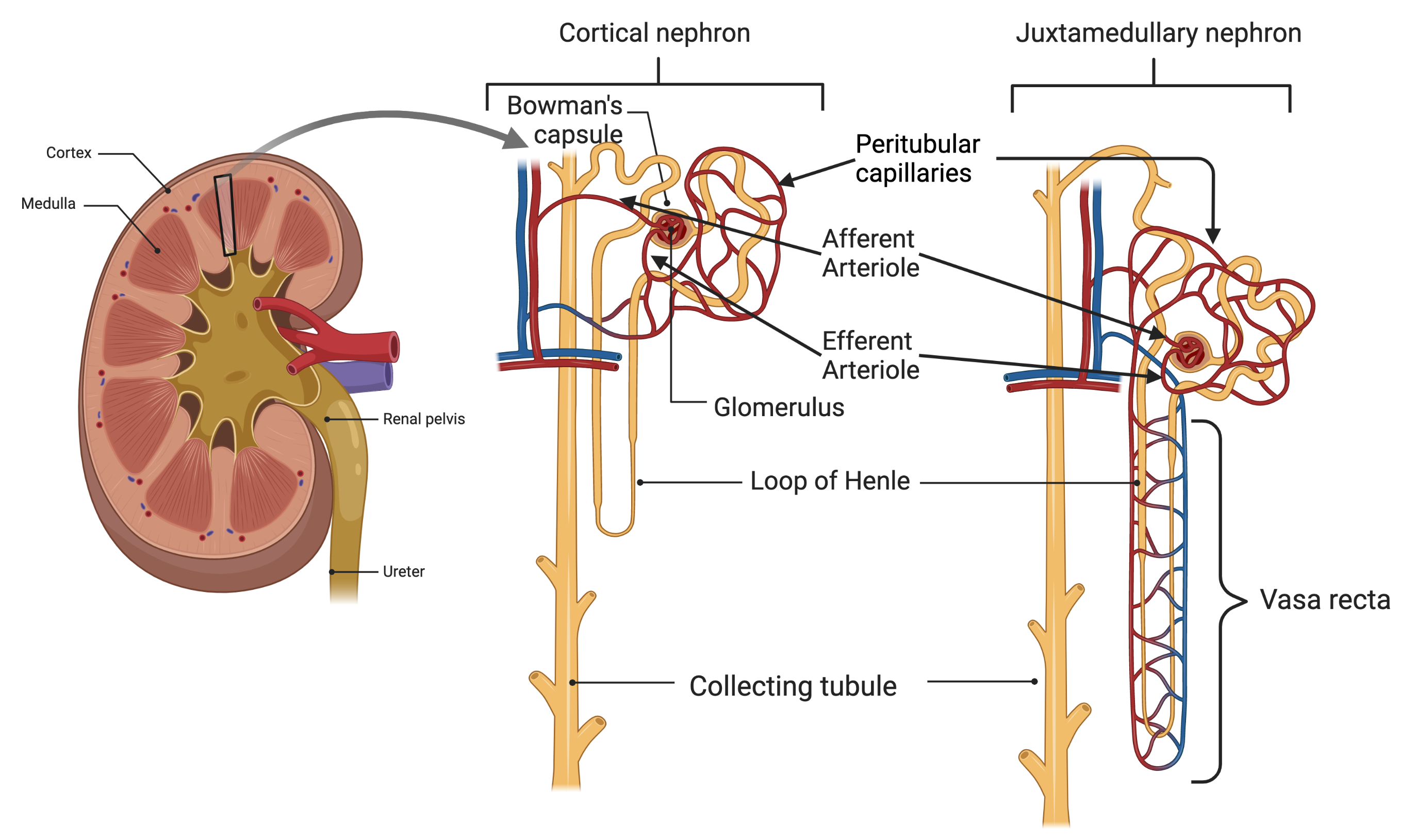
From this region the ultrafiltrate (which will become urine) travels through a looping tubular structure where the functions of re-absorption and secretion occurs. The segments of the nephron tubule that emerge from Bowman’s capsule includes several distinct functional units (in order): the proximal convoluted tubule, the proximal straight tubule, the loop of Henle (which contains a thin descending limb, a thin ascending limb, and a thick ascending limb), the distal convoluted tubule, and the collecting ducts. The distinct reabsorption and secretion functions across the tubule are based on a highly specialized epithelial cells lining each segment. The collecting ducts drain regionally into pouches which then drain larger pouches eventually reaching the renal pelvis which proceed into the ureter. Through this collecting duct system the urine from each kidney drains into a ureter and is transported to the bladder for storage and eventual elimination.
There are two types of nephrons, superficial cortical nephrons and juxtamedullary nephrons, which are distinguished by the location of their glomeruli and the length of their loops of Henle. The superficial cortical nephrons have their glomeruli in the outer cortex (which is more superficial) and have relatively short loops of Henle. The juxtamedullary nephrons have their glomeruli deeper in the kidney with larger glomeruli and therefore have higher glomerular filtration rates. The juxtamedullary nephrons also have long loops of Henle that go deep into the kidney that are essential for the concentration of urine.
Renal Blood Vessels
A unique aspect to renal blood vessels is that they have two sets of arterioles. Blood enters each kidney though the renal artery, which branches until the smallest arteries subdivide into the first set of arterioles, the afferent arterioles. The afferent arterioles function to deliver blood to the glomerular capillaries - a major site of filtration in the nephron. Following filtration blood travels through the second set of arterioles, the efferent arterioles, which deliver blood to the peritubular capillaries, a major site for water and solute reabsorption. Having arterioles before and after the glomerulus equips each nephron with a unique ability to regulate the hydrostatic pressure of the glomerular capillaries, resulting in changes to both renal blood flow and glomerular filtration rate. For example, constriction at either the afferent or efferent end of the glomerulus results in increased vascular resistance and reduced renal blood flow. To further assist in homeostatic regulation of water-solute balance, the juxtamedullary nephrons have specialized capillary loops called Vasa Recta. These blood vessels travel in a similar form to the Loop of Henle and act as osmotic exchanges, which regulate urine concentration.
8.2.2 Renal Clearance
Renal clearance is the volume of plasma cleared of a substance per unit time and is determined by filtration, reabsorption and secretion.
-
1.
Filtration (indiscriminate elimination): pushing filtrate (plasma) out of the capillary into Bowman’s capsule
-
2.
Reabsorption (selective): selectively pulling substances (including water) back into the capillary
-
3.
Secretion (selective): selectively pushing substances out of the capillary into the renal tubule
Substances with the highest clearances are both filtered and secreted and may be completely removed on a single pass of blood through the kidneys. Substances with the lowest clearances either are not filtered or are filtered and subsequently reabsorbed and may not be removed at all. Renal clearance is based on the rate substances are removed (cleared) from plasma in the kidneys. The higher the renal clearance of a substance, the more plasma that is cleared of the substance.
For example, kidneys regulate the renal clearance of to match what is absorbed so that people do not have to adjust what they ingest. In situations of reduced or impaired renal function dietary modification of may be necessary. The general principle is that normal physiology provides individuals lots of freedom since it can make adjustments to buffer the consequences of behavior. As that physiological capacity is lost with aging and various chronic conditions, behavior must be adjusted to avoid the consequences.
Renal Clearance of Substances
Renal clearance of albumin is approximately zero because albumin is not filtered. The renal clearance of glucose is also zero. Glucose is filtered and then completely reabsorbed. , urea, phosphate, and have clearances that are higher than zero because they are filtered and then partially, and selectively, reabsorbed as required. Inulin, a fructose sugar, is a special case that has made it valuable in the study of kidney function. Inulin is freely and completely filtered across the glomerular capillaries and it is neither reabsorbed nor secreted. Therefore the amount of inulin in the urine is the amount that was filtered. Its clearance measures the glomerular filtration rate (GFR) and has been utilized as the gold standard for identifying other useful measures of the GFR.
8.2.3 Urine Formation & Excretion by Micturition
Urine is formed by anything (including water) that is filtered and not reabsorbed, or not filtered and then secreted. Urine leaves the kidneys and flows through the ureters into the bladder. Peristaltic contractions of smooth muscles in the ureter, enhanced by parasympathetic activation, force urine from the kidneys toward the bladder which help maintain the flow of urine from the kidneys even as pressure rises in the bladder.
The bladder is a smooth muscle chamber with two parts. The bladder body collects urine and the neck is a funnel-shaped extension that connects with the urethra. The smooth muscle of the bladder can increase bladder pressure to 40-60 and play a major role in emptying the bladder during micturition.
The smooth muscle of the bladder wall is influenced by the autonomic nervous system and a spinal cord stretch reflex. Micturition (urination) is the process of emptying the bladder when it is filled. Filling of the bladder increases its pressure and therefore wall tension rises. Pelvic nerves connect with the spinal cord through the sacral plexus include sensory and motor nerve fibers. The sensory nerve fibers detect when the bladder wall tension rises above a threshold level that activates the micturition reflex to empty the bladder or, if this fails, creates a conscious desire to urinate. The motor nerves transmitted to the pelvic nerves are parasympathetic fibers.
Background: Urinary Semantics
The terms micturition and urination are equivalent and can be used interchangeably. However, the reflex that triggers urination is the micturition reflex. Though, if you heard someone say urination reflex, I’m quite sure you’d know what they meant.
The bladder neck includes the internal sphincter muscle interlaced with a large amount of elastic tissue. The natural tone of the internal sphincter muscle keeps the bladder from emptying until the pressure in the main part of the bladder rises above a critical threshold. Beyond the neck of the bladder the urethra passes through the urogenital diaphragm, which includes a layer of muscle called the external sphincter. This muscle is a voluntary skeletal muscle and is used to prevent urination even when the micturition reflex is attempting to empty the bladder.
Urinary Incontinence
There are numerous potential causes of urinary incontinence. A common cause of all forms includes the inability to prevent bladder pressure, either through the micturition reflex or external provocations such as abdominal pressure creating bladder pressure (for example during coughing, laughing, valsalva, etc), from resulting in urination. The micturition reflex includes a complete cycle of (1) progressive and rapid increase in bladder pressure, (2) sustained increase in bladder pressure, and (3) return of the pressure to the basal tone of the bladder. The steps include [hall_guyton_2020]:
-
1.
Sensory signals from the bladder wall stretch receptors are conducted to sacral segments of the spinal cord through the pelvic nerves and then reflexively back to the bladder through the parasympathetic nerves by way of the pelvic nerves.
-
2.
Once the micturition reflex is sufficiently powerful, it causes another reflex that passes through the pudendal nerves to the external sphincter to inhibit it. If this inhibition is more potent than the voluntary constrictor signals to the external sphincter, urination occurs.
-
3.
The micturition reflex is an autonomic spinal cord reflex, but it can be inhibited or facilitated by centers in the brain stem, mainly the pons, and several centers in the cerebral cortex that are mainly excitatory but can become inhibitory.
Not urinating includes the collaborative function of the internal and external sphincter. Both are involved in the micturition reflex above. Voluntary tone of the external sphincter must be properly timed and sufficient to prevent urination when the micturition reflex is sufficiently powerful. The strength of the micturition reflex is influenced by bladder pressure and wall tension. Therefore bladder pressure can be increased by urine volume or increased abdominal pressure (for example, that comes on during laughing, sneezing or coughing). Much of Women’s Health Physical Therapy is centered on helping post-partum women regain voluntary control over the micturition reflex. It is important to point out that the terminology ”voluntary” simply means a voluntary skeletal muscle. It does not necessarily mean conscious control. For example, several voluntary skeletal muscles are involved when you breath, maintain a particular posture or walk. While being voluntary they are not necessarily conscious (you can think about your breathing, posture and walking muscle activation, but you typically do not).
8.3 Filtration-Reabsorption-Secretion
8.3.1 Filtration
Glomerular filtration is the first step in the formation of urine. The rate of glomerular filtration is appropriately called the glomerular filtration rate (GFR). As blood enters the glomerular capillaries, a portion of that blood is filtered into Bowman’s capsule. The fluid that is filtered is similar to interstitial fluid and at this point is called an ultrafiltrate. Ultrafiltrate contains water and all of the small solutes of blood, but not proteins and blood cells. The pressures responsible for glomerular filtration are similar to the pressures operating for micro-circulation filtration in systemic capillaries (Chapter 7). However, the characteristics and surface area of glomerular capillaries result in a much higher GFR than filtration in systemic capillaries (glomerular capillaries have a higher filtration coefficient than systemic circulation capillaries). The ultrafiltrate of glomerular filtration renters the peritubular capillaries through reabsorption and can be more selective. The ultrafiltrate that does not re-enter the vascular fluid does not enter lymph vessels, but rather proceeds through the renal tubules for urine formation and excretion.
Factors Influencing Glomerular Filtration
As with microcirculation there are four pressures for filtration: two hydrostatic pressures and two osmotic pressures. Applying these pressures to glomerular capillaries, there is one small modification because the osmotic pressure of Bowman’s capsule is considered to be zero and therefore is not considered for GFR [costanzo_physiology_2013]. The primary factors that influence GFR include:
-
1.
Glomerular filtration coefficient
-
2.
Hydrostatic pressure in glomerular capillary
-
3.
Hydrostatic pressure in Bowman’s capsule
-
4.
Osmotic pressure in glomerular capillary
-
5.
Net filtration pressure (nfp)
Glomerular filtration coefficient
The glomerular filtration coefficient is the permeability of the glomerular capillary wall. Two factors that contribute to filtration coefficient are the water permeability per unit of surface area and the total surface area. The coefficient for glomerular capillaries is approximately 100-fold greater than systemic capillaries (such as skeletal muscle capillaries) because of the combination of a higher total surface area and a higher intrinsic water permeability. The consequence of this extremely high coefficient is that much more fluid is filtered from glomerular capillaries than from other capillaries. This coefficient can be decreased with damage to the glomerular capillaries brought on by chronic conditions such as diabetes and high blood pressure (hypertension (HTN)).
Hydrostatic pressure in glomerular capillary (favors filtration)
The hydrostatic pressure in glomerular capillary favors filtration. Compared with systemic capillaries, it is relatively high (60 ). In systemic capillaries, hydrostatic pressure falls along the length of the capillary; in glomerular capillaries, it remains constant along the entire length. This is possible because it is regulated with two sets of arterioles, those entering the glomerular capillaries (afferent arterioles) and those exiting the glomerular capillaries (the efferent arterioles). This regulatory system also allows for it to remain relatively constant across a wide range of blood pressures (80-200 ). Changes in the hydrostatic pressure in glomerular capillary are produced by changes in the resistance of the afferent and efferent arterioles which subsequently change renal blood flow (RBF). Changes in GFR depend on changes to to this hydrostatic pressure which depend on both the afferent and efferent arterioles.
Hydrostatic pressure in Bowman’s capsule (opposes filtration)
The hydrostatic pressure in Bowman’s capsule is a pressure opposing filtration. The origin of this pressure (18 ) is the fluid present in Bowman’s capsule and the tubule of the nephron. Changes in this pressure can be produced by obstructing urine flow (e.g., ureter stone or constriction). If the ureter is constricted, urine cannot flow through that ureter to the bladder, causing urine to back up in the kidney. Consequently, hydrostatic pressure in the nephrons will increase as far back as Bowman’s capsule. An increase in the hydrostatic pressure in Bowman’s capsule decreases the GFR.
Osmotic pressure in glomerular capillary (opposes filtration)
The osmotic pressure in glomerular capillary is a pressure opposing filtration. It is determined primarily by the protein concentration of glomerular capillary blood. It progressively increases along the capillary length as fluid (but not protein) is filtered out of the capillary. The osmotic pressure in glomerular capillary eventually increases to the point where net filtration pressure becomes zero and glomerular filtration stops (filtration equilibrium). Changes in the osmotic pressure in glomerular capillary are produced by changes in plasma protein concentration. Increases in plasma protein concentration produce increases, which decrease the GFR. Decreases in plasma protein concentration produce decreases in osmotic pressure in glomerular capillary which increase GFR.
Net filtration pressure
The net filtration pressure (nfp) is the simple sum of of the previous three pressures with those favoring filtration as positive numbers, and those opposing filtration as negative numbers. The nfp in the glomerular capillaries is positive, and thus favors filtration. The direction of fluid movement is always out of the capillaries. The greater the nfp, the higher the GFR (they are directly related).
Clinical Estimation of GFR
The clinical estimation of GFR provides an overall assessment of renal function. Inulin is freely and completely filtered across the glomerular capillaries and it is neither reabsorbed nor secreted. The amount of inulin in the urine is the amount that was filtered and its clearance measures the GFR. Inulin is the gold standard for identifying other useful measures of the GFR. The use of inulin to measure GFR requires a prolonged highly controlled situation that includes set infusion of inulin and collection of urine for hours to record its clearance. Therefore, inulin is rarely utilized clinically to measure GFR.
BUN and creatinine
The closest endogenous111Originating or produced within an organism, tissue, or cell. substance to inulin for estimation of GFR is is creatinine. It is freely filtered across the glomerular capillaries but is also secreted to a small extent. Therefore, clearance of creatinine slightly overestimates the GFR. However, since creatinine is an endogenous substance it does not need to be infused in order to estimate GFR. Urea is another endogenous substance that does not need to be infused that is filtered across the glomerular capillaries. Together, blood urea nitrogen (BUN) and serum creatinine concentration are used to estimate GFR because each substance depends on the filtration step in order to be excreted in urine; urea is reabsorbed in small quantities, and creatinine is secreted in small quantities.
With a decrease in GFR due to renal conditions (such as renal failure), BUN and serum creatinine both increase because they are not adequately filtered. But with lower blood volume (hypovolemia, due to dehydration) or reduced RBF (perfusion, due to heart failure) there is also decreased GFR and both BUN and serum creatinine are increased. However, because urea is reabsorbed and creatinine is not, BUN increases more than serum creatinine. Therefore an indicator of hypovolemia or heart failure caused renal insufficiency is an increased ratio of BUN/creatinine to more than 20. While renal failure due to renal causes produces an increase in both BUN and serum creatinine, it does not produce an increase in the BUN/creatinine ratio [hall_guyton_2020].
| BUN | Creatinine | BUN/Creatinine Ratio | |
|---|---|---|---|
| Renal Disease (renal failure) | Normal | ||
| Renal Blood Flow (hypovolemia, heart failure) |
8.3.2 Tubular Reabsorption
Glomerular filtration results in the production of large quantities of ultrafiltrate each day (approximately 180 L/day). If all of this ultrafiltrate were excreted as urine the following quantities would be lost each day: 180 of water; 25,200 of ; 19,800 of ; 4320 of ; and 14,400 of glucose. Each of these losses is 10 times more than the amount present in the entire ECF. This is clearly not a sustainable approach to mass balance.
There are a set of reabsorption mechanisms in the epithelial cells lining the renal tubule that return these (and other) substances to the peritubular capillaries. The details of these mechanisms are beyond the scope of this text. Overall, reabsorption includes the return to capillaries through several different transport proteins that either selectively and directly transport substances, or through the transport of substances which then manipulate the osmolarity between the renal tubule and the peritubular capillaries. Water and many solutes (, , , glucose, amino acids, urea, , , phosphate, lactate, and citrate) are reabsorbed from ultrafiltrate back into the peritubular capillaries. If reabsorption did not occur, most of these constituents of ECF would be rapidly lost in the urine.
This approach, the generation of a large quantity of ultrafiltrate and the selective reabsorption back into the capillaries, is a highly efficient way to filter and clear waste products (urea, creatinine that have limited reabsorption), and regulate the blood and ECF content of other substances. For example, even though glucose is filtered and then reabsorbed, in certain hyperglycemic situations with diabetes, glucose cannot be fully reabsorbed which results in glucose in the urine (glycosuria). Glycosuria contributes to an upper limit of blood glucose and its acutely deleterious effects, but it is not a healthy long term homeostatic mechanism for glucose control.
Reabsorption is adjusted based on ECF fluid volume. Increased ECF volume inhibits tubule reabsorption which results in loss of ECF fluid volume, and decreased ECF volume stimulates tubule reabsorption which results in an increase in ECF. Different diuretics (drugs that promote water removal in urine) work at different points along the renal tubule to reduce reabsorption of water.
8.3.3 Tubular Secretion
Secretion mechanisms in the epithelial cells can remove selected and specific substances from the peritubular capillary blood and add them to urine. Organic acids, organic bases, and are secreted from peritubular capillary blood into tubular fluid as needed. In addition to filtration, secretion provides a mechanism for excreting substances in the urine. Compared to filtration secretion is selective like reabsorption. The secretion mechanisms involve transporters in the membranes of the epithelial cells lining the renal tubule.
8.3.4 Excretion Rate: putting it all together
Excretion rate refers to the amount of a substance excreted per unit time and is related to the clearance rate. However the excretion rate is in terms of the amount of a substance excreted per time frame () whereas the clearance rate is in terms of the volume of plasma cleared of the substances (). The overall reabsorption or secretion rate of a substance is the difference between the amount filtered and its excretion rate. The excretion rate reflects the overall result of glomerular filtration, tubular reabsorption, and tubular secretion. It can be compared with the amount filtered to determine whether a substance has been reabsorbed or secreted. The difference between the amount filtered and the excretion rate is the rate of net reabsorption or net secretion. When the amount filtered is greater than the excretion rate, there has been reabsorption of the substance. If the amount filtered is less than the excretion rate, there has been secretion of the substance.
8.4 Renal Regulation
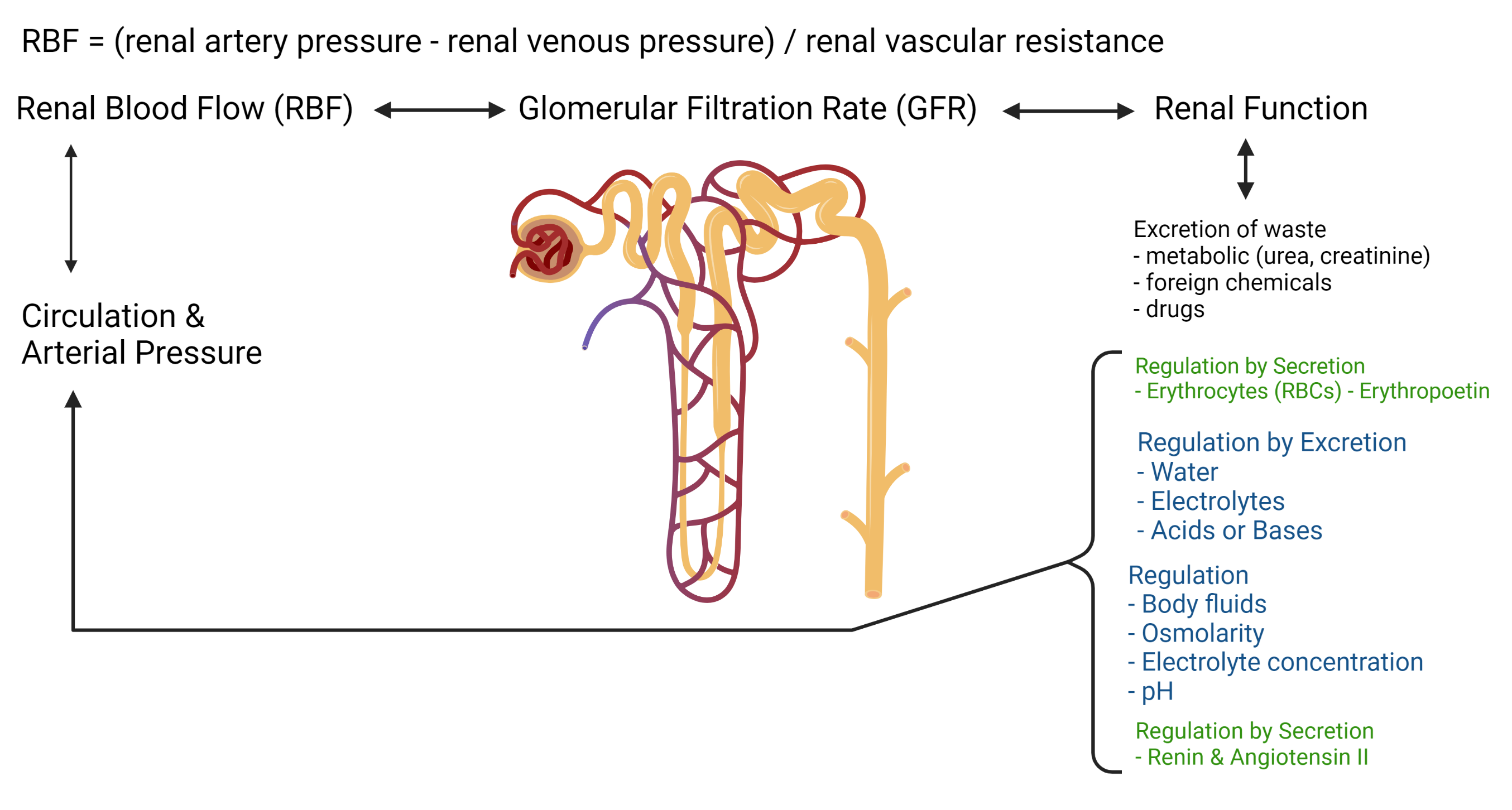
Renal regulation includes the regulation of renal function, and the renal regulation of several critical characteristics of ECF. These two aspects of regulation are integrated and coordinated. Renal function regulates blood volume and osmolarity by adjusting both water content and electrolytes (primarily ). Regulation of content is balanced with regulation of concentration. There are limits to what the kidneys do to adjust water volume and osmolarity because they are also making sure the concentration stays within fairly tight limits. The regulation of renal function is similarly based on alterations in blood volume that influence glomerular filtration, tubular reabsorption and tubular secretion. Figure 8.5 depicts the bidirectional relationship between renal blood flow (RBF), GFR and renal function (excretion rates and other functions). Renal function influences circulation and arterial pressure which then influences RBF. Arterial pressure is also regulated and is integrated with renal regulation via the hormones renin, angiotensin II, aldosterone and anti-diuretic hormone (ADH).
8.4.1 Sodium
The kidneys maintain a normal body content by ensuring that excretion equals intake, a matching process called balance. balance is achieved by variations in the reabsorption of . If excretion is less than intake, then there is positive balance. Extra is retained, primarily in the ECF. When the content of ECF is increased, there is increased ECF osmolarity and thus volume; blood volume and arterial pressure also increase, and there may be edema. If excretion is greater than intake, then a person is in negative balance. When excess is lost from the body, there is a decreased content of ECF, decreased ECF osmolarity and volume, increased ICF volume, and decreased blood volume and arterial pressure.
8.4.2 Potassium
balance is maintained by shifts of across cell membranes and by renal regulation. The renal mechanisms for balance include filtration, reabsorption, and secretion. Secretion is influenced by dietary , aldosterone, acid-base balance, and flow rate. With low intake, more is reabsorbed and less is secreted; with high intake less is reabsorbed and more is secreted. The secretion of in the distal tubule and collecting ducts allows the fine-tuning of excretion to maintain balance.
Acid-base disturbances can effect blood concentration due to alterations in secretion. Alkalosis increases secretion, and acidosis decreases secretion. Therefore, conditions such as chronic pulmonary disease that increase blood carbon dioxide and decrease blood pH (respiratory acidosis) can be accompanied by hyperkalemia.
Commonly used diuretics, the loop diuretics and the thiazide diuretics, cause increased excretion. Therefore, an important side effect of diuretic therapy is hypokalemia. The -sparing diuretics (e.g., spironolactone, amiloride, triamterene) do not cause increased excretion because they inhibit all of the actions of aldosterone and inhibit secretion. The most common use of -sparing diuretics is in combination with the loop or thiazide diuretics to offset hypokalemia produced by those drugs.
8.4.3 Calcium
Calcium regulation involves kidney reabsorption in response to parathyroid hormone. The overall coordinated process of calcium regulation involves variations in intestinal absorption and release from bones. Therefore, it is best considered with an understanding of intestinal absorption.
8.4.4 Acid Base Balance
There are three ways the kidneys influence acid base balance. First, the secretion of ; second the reabsorption of ; and third, the production of new . It is best to consider kidney (metabolic) causes and compensations of acid-base balance along with respiratory causes and compensations of acid-base balance. Therefore, clinical interpretation of acid-base imbalance is in the chapter on respiration.
8.4.5 Blood Volume & Osmolarity
Blood volume is regulated based on two primary factors: 1. impact on BP, RBF and subsequently GFR; 2. impact of water in plasma on ECF osmolarity. These are not completely separate regulatory pathways (there is overlap). The overlap is clear when considering the integration of the hormonal mechanisms underlying the regulation of blood pressure and osmolarity. The primary hormones to be considered include: renin, angiotensin II, anti-diuretic hormone (ADH) and aldosterone. The influence of these three hormones on renal function is summarized in Table 8.2. How these hormones and renal function is involved in long term blood pressure regulation is covered in Chapter 9 on Circulation.
| Hormone | Activated by | Effects |
|---|---|---|
| Renin | GFR | Angiotensin Converting Enzyme (ACE) |
| Angiotensin II | Angiotensin Converting Enzyme (ACE) | Aldosterone; reabsorption, GFR |
| Aldosterone | Angiotensin II & plasma concentration | secretion, reabsorption |
| Anti-Diuretic Hormone (ADH) | plasma osmolarity | reabsorption |
Water Balance
The intentional and incidental loss of water requires regular ingestion and absorption of water. Situations with more water loss than intake is a negative water balance. Situations with more water intake than loss is a positive water balance.
| (8.1) |
Monitoring water balance occurs through the monitoring of plasma osmolarity by the anterior hypothalamus (osmoreceptors). The response to changes in osmolarity involves ADH which has multiple end organ effects, including those that influence perception (thirst) and behavior (drinking).
Response to Negative Water Balance
-
1.
Water is continuously lost from the body in sweat and in water vapor from the mouth and nose, as well as in urine (the kidneys must continue making urine to for waste excretion). If water is not replaced by drinking water, then plasma osmolarity increases.
-
2.
Increased plasma osmolarity stimulates osmoreceptors in the anterior hypothalamus, which are stimulated by changes in osmolarity of less than 1 mOsm/L. Stimulation of the hypothalamic osmoreceptors has two effects. It stimulates thirst, which drives drinking behavior. It also stimulates secretion of ADH from the posterior pituitary gland.
-
3.
The posterior pituitary gland secretes ADH. ADH circulates in the blood to the kidneys, where it produces increased water reabsorption. As more water is reabsorbed urine osmolarity increases and urine volume decreases (concentrated urine).
-
4.
Increased water reabsorption means that more water is returned to the body fluids. Coupled with increased thirst and drinking behavior, plasma osmolarity is decreased.
The overall response is an excellent example of coordinated homeostatic negative feedback. The original disturbance (increased plasma osmolarity) causes a set of feedback responses (thirst, secretion of ADH and increased water reabsorption) that decrease plasma osmolarity [hall_guyton_2020]. Negative water balance results in concentrated urine.
Response to Positive Water Balance
-
1.
Ingested water is distributed throughout the body fluids. Because the amount of solute in the body is unchanged, the added water dilutes the body fluids and cause a decrease in plasma osmolarity.
-
2.
The decrease in plasma osmolarity inhibits osmoreceptors in the anterior hypothalamus.
-
3.
Inhibition of the osmoreceptors has two effects. It decreases thirst and suppresses water drinking behavior. It also inhibits secretion of ADH from the posterior pituitary gland.
-
4.
When ADH secretion is inhibited, circulating levels of ADH are reduced and less ADH is delivered to the kidneys. Lower ADH levels decreases water reabsorption and water is excreted, decreasing urine osmolarity and increasing urine volume (diluted urine).
-
5.
Because less water is reabsorbed, less water is returned to the circulation. Coupled with the inhibition of thirst and the suppression of water drinking, plasma osmolarity increases back toward the normal value.
The overall response is an excellent example of coordinated homeostatic negative feedback. The original disturbance (decreased plasma osmolarity) causes a set of feedback responses (decreased thirst, less secretion of ADH and decreased water reabsorption) that increase plasma osmolarity [hall_guyton_2020]. Positive water balance results in diluted urine.
Electrolyte Induced Changes to Osmolarity
Water balance is not the only change that alters osmolarity and trigger the osmoreceptor - ADH response. Changes to osmolarity due to changes in solute concentrations also trigger the osmoreceptor - ADH response. The response coordinated by ADH primarily changes water content to adjust osmolarity, but also facilitates correction of solute concentrations, though not solute content. For example, if content increases with no change in water then concentration increases along with plasma osmolarity. This hypernatremia induced increase in osmolarity causes a shift in water out of the ICF, which partially reduces but does not normalize the ECF osmolarity. The still higher than normal osmolarity activates osmoreceptors that result in the release of ADH. ADH results in thirst, increased intake of water, and reduced loss of water. Together this response dilutes the plasma so that concentration is reduced along with osmolarity. A consequence is higher plasma volume and therefore higher blood volume, which may result in higher blood pressure. Ideally the kidneys continue to correct this problem with less reabsorption that returns plasma volume to a normal range.
8.5 Disturbances in Fluid Volume & Osmolarity
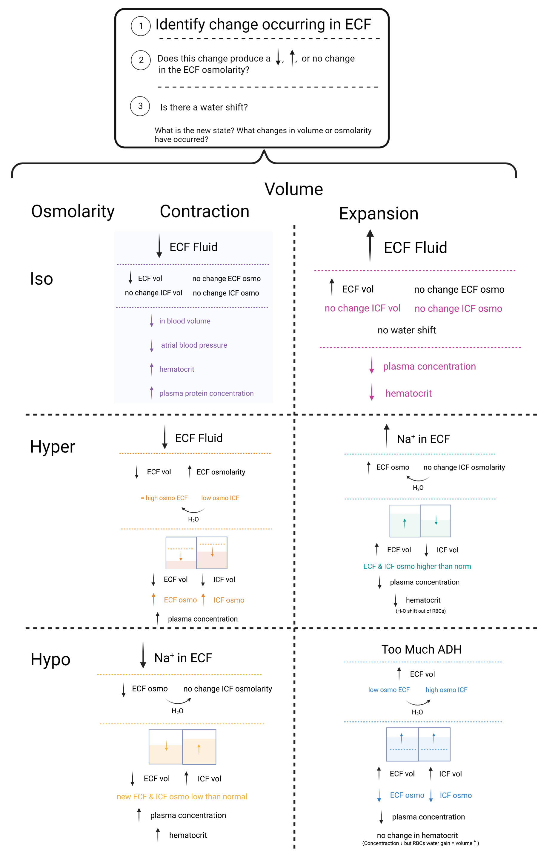
It is a useful exercise to understand and reason through disturbances in fluid volume (2) and osmolarity (3). Altogether six disturbances. It is not intended that readers would memorize the sequences below, but rather understand them and, if needed, recreate them from thinking through the processes involved from an understanding of the principles. The descriptions below do not include all of the possible trajectories or all of the physiological responses. One goal is to start considering the impact of ECF volume on hematocrit, and the fact that the volume of a RBC is intracellular.
Osmolarity is the concentration of osmotically active particles, expressed as milliosmoles per liter (). The normal value for osmolarity of the body fluids is approximately 290 in both ECF and ICF since water will shift between these spaces until osmolarity equilibrates [costanzo_physiology_2013].
Plasma osmolarity () can be estimated from the plasma concentration (), plasma glucose concentration (), and blood urea nitrogen (BUN) (), since these are the major solutes of ECF and plasma.
| (8.2) |
The concentration is multiplied by 2 because is balanced by an equal concentration of negative ions (in plasma, these anions are and .)222Despite common assumptions that the outside of the cell is positive and the inside of the cell is negative, there is electroneutrality in both ECF and ICF fluid. Recall from Chapter 4 that membrane potentials are generated from the movement of the ions, not the concentration of the ions. The glucose concentration in mg/dL is converted to mOsm/L when it is divided by 18. The BUN in mg/dL is converted to mOsm/L when it is divided by 2.8. Calculating Equation 8.2 with the values of 140 of , 80 of glucose, and 15 for BUN results in 289.79 .
In the following sections volume contraction means a decrease in ECF volume. Volume expansion means an increase in ECF volume. In place of the more general terms isotonic, hypertonic and hypotonic the more specific terms isosmotic, hyperosmotic, and hyposmotic refer to the osmolarity of the ECF for the osmolarity disturbances. Consistent with Chapter 7, an isosmotic disturbance means that there is no change in ECF osmolarity; a hyperosmotic disturbance means that there has been an increase in ECF osmolarity; and a hyposmotic disturbance means that there has been a decrease in ECF osmolarity.
Three-Step Approach to Reasoning Through Disturbances in Fluid Volume & Osmolarity
To understand these disturbances, a three-step approach is recommended ([costanzo_physiology_2013]):
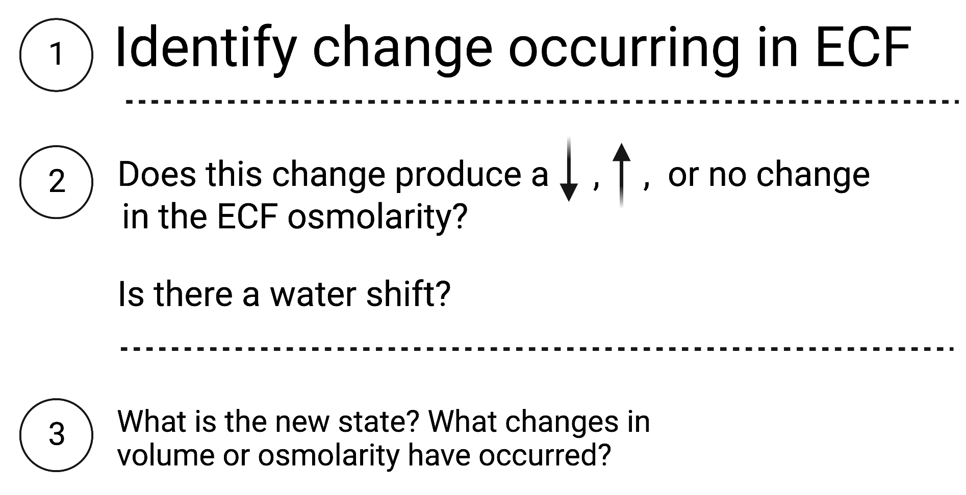
Six Disturbances: Volume x Osmolarity
The following six pages each includes a description of a volume osmolarity disturbance. Each page also includes a graphic depiction of how to examine each using the three step approach.
| Osmolarity | Volume Contraction | Volume Expansion |
|---|---|---|
| Isosmotic | Diarrhea | Infusion of NaCl |
| Hyperosmotic | Sweating Lactate | High NaCl Intake |
| Hyposmotic | Adrenal Insufficiency | SIADH |
8.5.1 Isosmotic Volume Contraction: Diarrhea
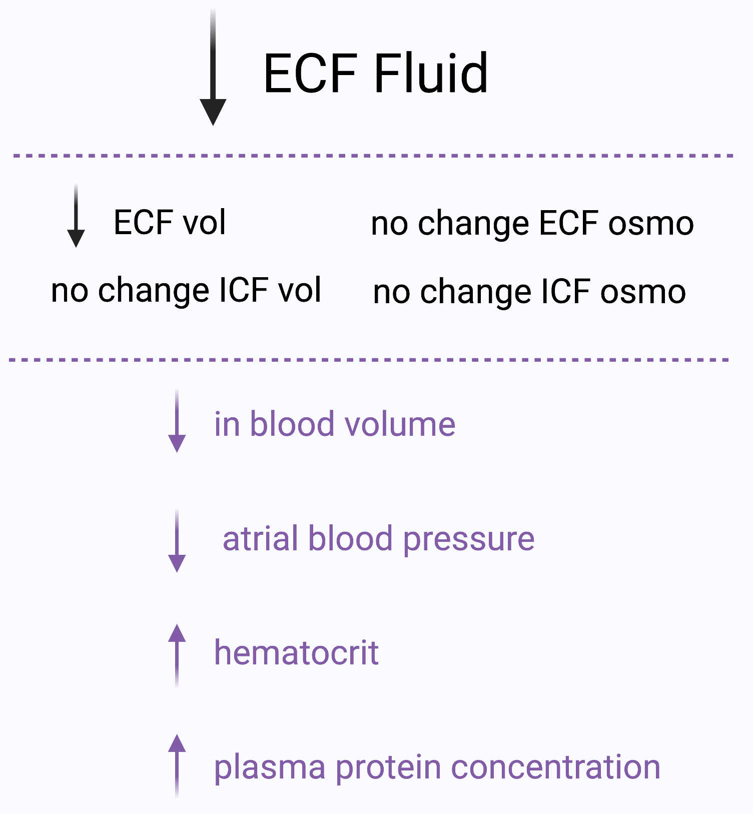
Diarrhea results in a large volume loss of fluid from the gastrointestinal tract. The osmolarity of the fluid lost is approximately equal to that of the ECF (isosmotic). ECF volume decreases, but there is no accompanying change in ECF osmolarity. Therefore there is no need for a fluid shift across cell membranes and ICF volume remains unchanged. In the new steady state, ECF volume decreases and the osmolarities of ECF and ICF are unchanged. The decrease in ECF volume means that blood volume (a component of ECF) also is reduced, which produces a decrease in arterial pressure. Other consequences of diarrhea include increased hematocrit and increased plasma protein concentration.
8.5.2 Hyperosmotic Volume Contraction: Sweating
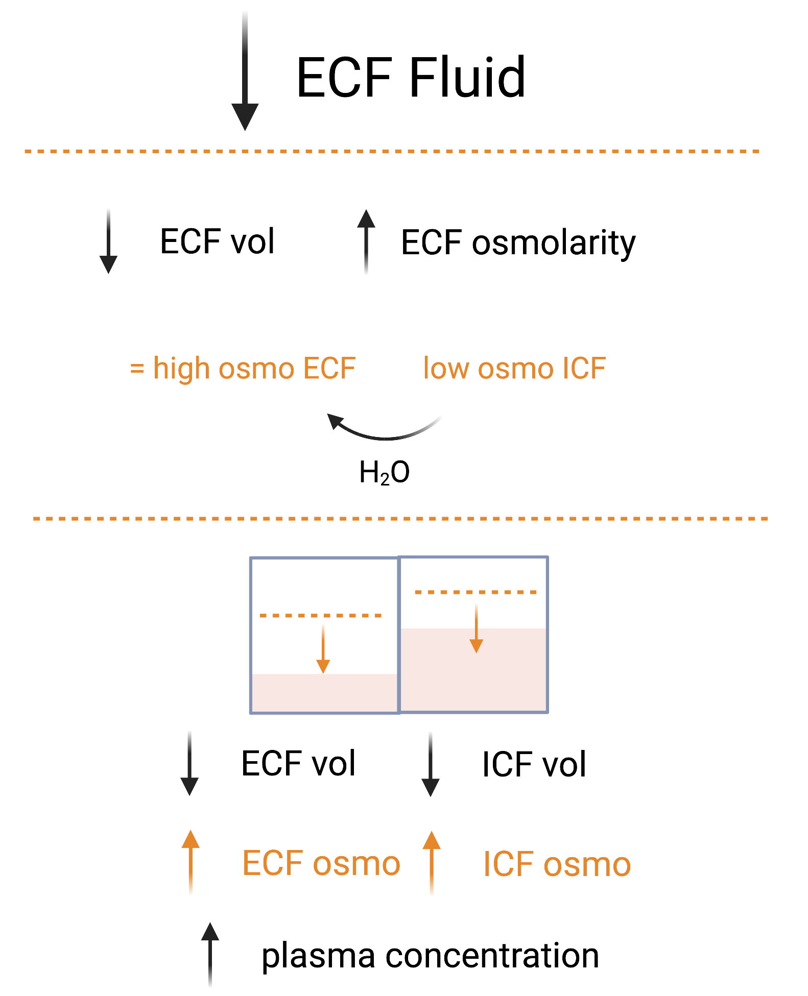
Water deprivation (negative mass balance of water) in situations with sweating (hot environments and/or high metabolism) results in net losses to both NaCl and water in sweat. Sweat is hyposmotic relative to ECF. Sweat contains relatively more water than solute. When sweating hyposmotic fluid is lost from the ECF. ECF volume decreases and ECF osmolarity increases. ECF osmolarity is transiently higher than ICF osmolarity, and this difference in osmolarity causes water to shift from ICF into ECF until ICF and ECF osmolarity equalizes. In the new steady state, both ECF and ICF volumes are decreased and ECF and ICF osmolarities increased. In hyperosmotic volume contraction, the plasma protein concentration is increased but the HcT is unchanged. The explanation for the increase in plasma protein concentration is straightforward: Fluid is lost from ECF, and the plasma protein remaining behind becomes concentrated. It is less obvious, however, why the hematocrit is unchanged. Loss of fluid from ECF alone would cause an increase in the concentration of red blood cells and an increase in HcT. However, there also is a fluid shift in this disturbance: Water moves from ICF to ECF. Because RBCs are cells, water shifts out of them, decreasing their volume. Thus, the concentration of red blood cells increases, but red blood cell volume decreases. The two effects offset each other, and HcT is unchanged. The shift of water from ICF to ECF offsets the loss of ECF from water loss.
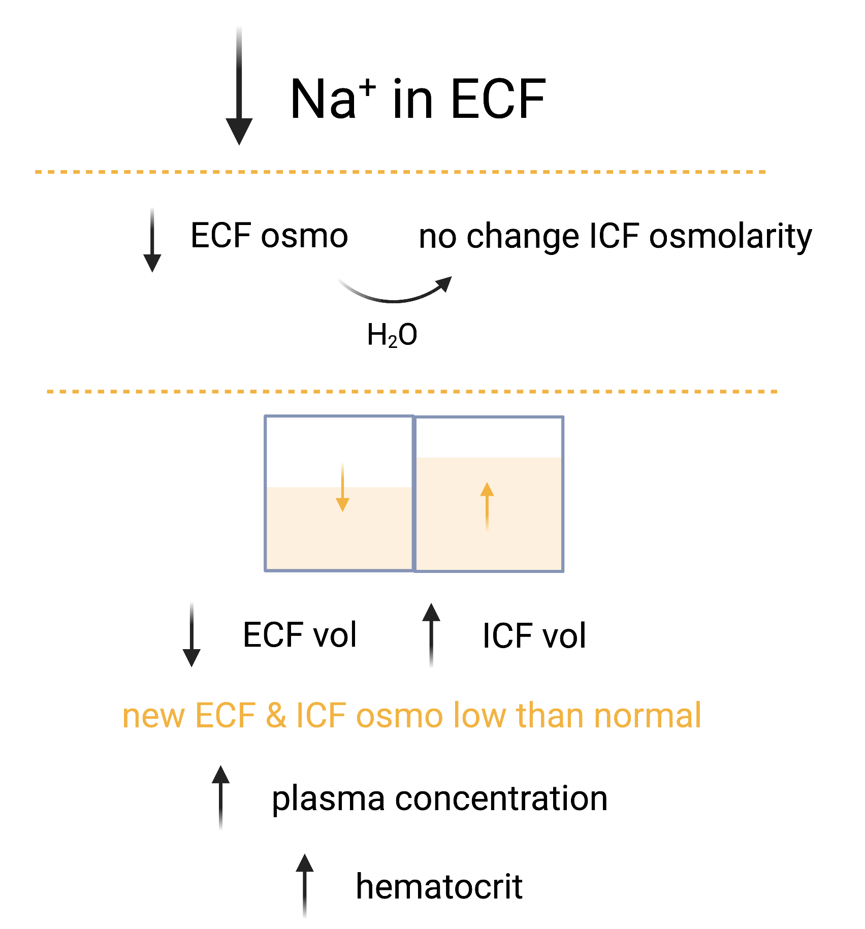
8.5.3 Hyposmotic Volume Contraction: Adrenal Insufficiency
Adrenal insufficiency includes a deficiency in aldosterone, a hormone that promotes reabsorption. Aldosterone deficiency results in excess NaCl excreted in the urine. Because NaCl is an ECF solute, ECF osmolarity decreases. ECF osmolarity is less than ICF osmolarity and causes water to shift from ECF to ICF until osmolarity equilibrium. In the new steady state, both ECF and ICF osmolarities are lower than normal. The shift of water results in decreased ECF volume and increased ICF volume. In hyposmotic volume contraction, both plasma protein concentration and HcT will be increased because of the decrease in ECF volume. HcT also increases because of the shift of water into red blood cells, increasing cell volume.
8.5.4 Isosmotic Volume Expansion: Infusion of NaCl
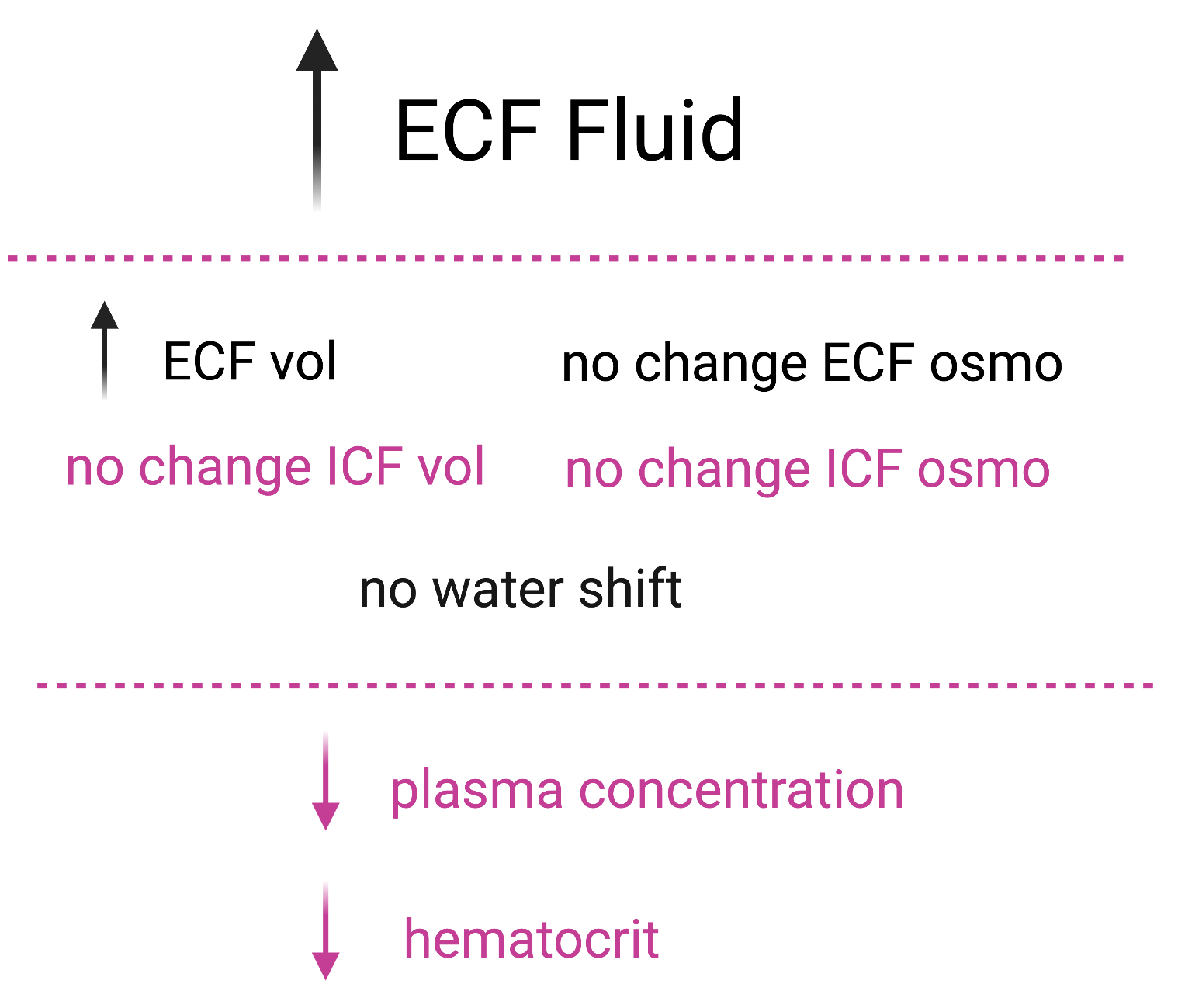
An infusion of isotonic NaCl presents the opposite clinical picture of losing isotonic fluid through diarrhea. Because NaCl is an extracellular solute, all isotonic NaCl solution is added to the ECF, causing an increase in ECF volume but no change in ECF osmolarity. There is no shift of water between ICF and ECF because there is no difference in osmolarity between the two compartments. Both plasma protein concentration and HcT decrease because of the increase in ECF volume.
8.5.5 Hyperosmotic Volume Expansion: High NaCl Intake
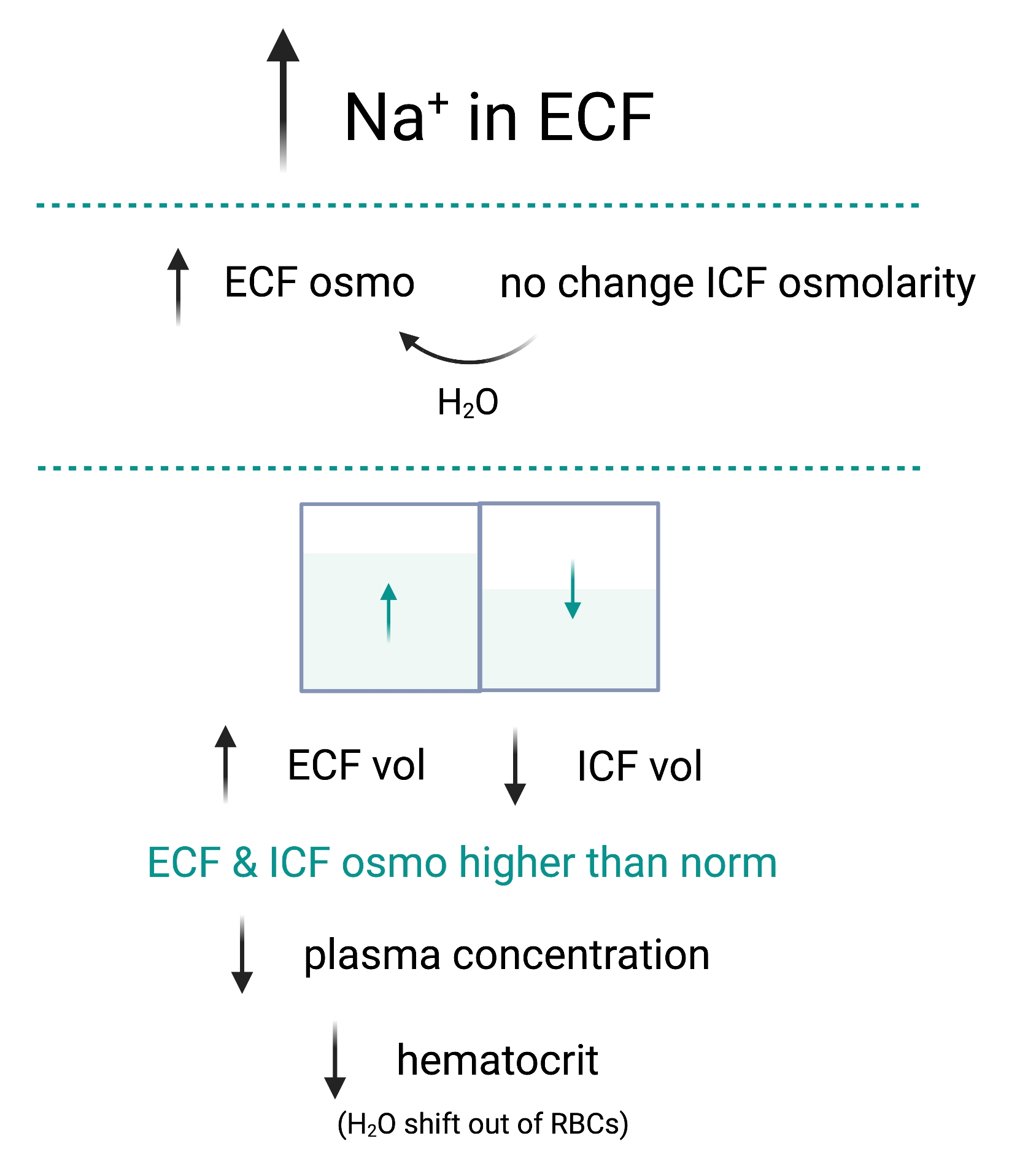
Ingesting dry NaCl (for example, eating a salty snack) increases the total amount of in the ECF and osmolarity increases transiently as the higher ECF osmolarity causes water to shift from ICF to ECF, decreasing ICF volume and increasing ECF volume. In the new steady state, both ECF and ICF osmolarities are higher than normal and equal to each other. Because of the shift of water out of cells, ICF volume will decrease and ECF volume will increase. In hyperosmotic volume expansion, both plasma protein concentration and hematocrit will decrease due to the increase in ECF volume. Hematocrit also will be decreased because of the water shift out of the red blood cells.
8.5.6 Hyposmotic Volume Expansion: SIADH
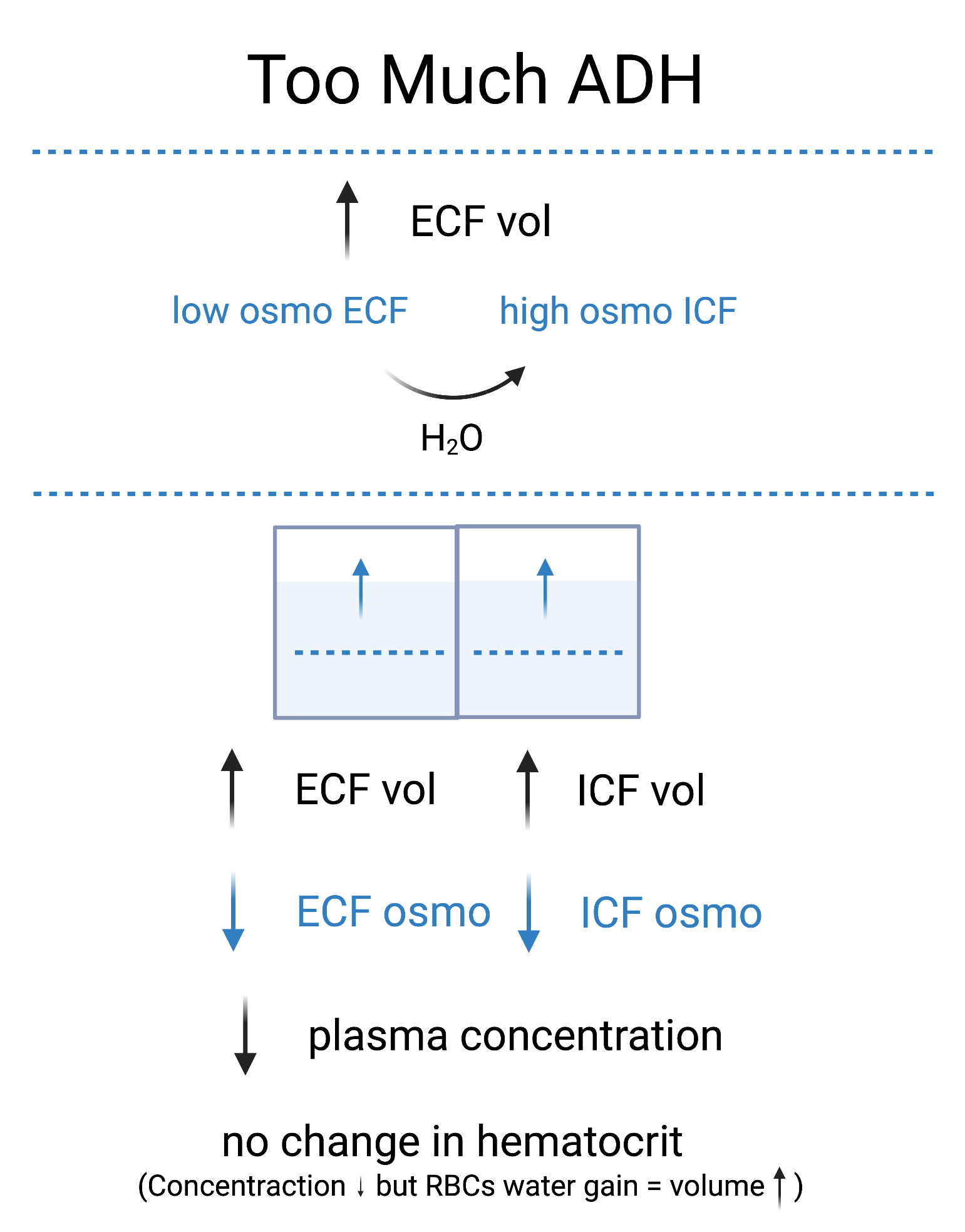
Syndrome of inappropriate antidiuretic hormone (SIADH) secretes inappropriately high levels of ADH, which promotes too much water reabsorption. The excess water is retained and distributed throughout the total body water. ECF is diluted and transiently has a lower osmolarity. Water shifts into the ICF since it has higher osmolarity. When compared with the normal state, ECF and ICF volumes are both increased and ECF and ICF osmolarities will be decreased. In hyposmotic volume expansion, plasma protein concentration is decreased by dilution. The hematocrit is unchanged as a result of two offsetting effects: The concentration of red blood cells decreases because of dilution, but RBC volume increases because water shifts into the cells. But this increase in RBC volume (due to water) does not necessarily increase oxygen carrying capacity of HbG so maintaining HcT in the situation of hyposmotic volume expansion does not necessarily mean oxygen carrying capacity is normal.
8.5.7 Summary of Disturbances in Fluid Volume & Osmolarity
8.6 Muscle Connections
8.6.1 Speculation on Water Homeostasis in Muscle Function & Frailty
Older adults have reduced thirst sensation and the ability to concentrate urine, together this results in increased ECF osmolarity (hyperosmotic stress) [lorenzo_role_2019]. Hyperosmotic stress leads to cell dehydration (reduced ICF), which has consequences for the intracellular protein structure and function. In severe situations this can result in cell damage. There is also some evidence to suggest that cell volume may act as a signal, with cell swelling acting as an anabolic signal (promotes hypertrophy) and cell shrinkage acting as a catabolic signal (promotes atrophy). One possible reason, or contributing factor, for the age related progressive loss in muscle mass and strength may be impaired ECF - ICF water homeostasis that originates from mild kidney insufficiency and a loss of thirst. Intracellular water content in lean mass has been related to muscle strength, functional capacity, and frailty risk, and has been proposed as an indicator of muscle quality and cell hydration [lorenzo_role_2019]. Muscle weakness can promote further decreases in fluid (and nutrient) intake due to challenges in ADLs. This can occur directly with difficulty attaining the necessary muscle tension for performing ADLs (either basic ADLs or instrumental ADLs), or with the accumulation of objective fatigue (difficulty sustaining ADLs)[collins_heart_2015]. Several chronic conditions that include renal insufficiency such as diabetes, high blood pressure, chronic heart failure, obstructive pulmonary disease and renal failure all share a common feature of muscle catabolism, atrophy and weakness. With these conditions the atrophy and weakness seem to go beyond disuse atrophy [adams_skeletal_2006]. A common, and plausible, feature of these conditions may be water homeostasis.
8.6.2 Rhabdomyolosis
Rhabdomyolosis is a condition in which damaged skeletal muscle breaks down rapidly. Symptoms include muscle pain; signs include weakness, muscle stiffness and edema. Signs may also include tea-colored urine, an irregular heartbeat, vomiting, and confusion. A positive urine myoglobin test provides supportive evidence. Myoglobin is harmful to nephrons so if rhabdomyolysis is severe it may lead to kidney failure. Common causes of muscle damage include crush injury, strenuous exercise, medications (including statins for lowering blood cholesterol), or substance abuse. Less common causes include infections, electrical injury, heat stroke, prolonged immobilization, lack of blood flow to a limb, or snake bites.
8.7 Summary
Mass balance is an essential concept for physical therapy practice. The need for intake and the intentional or incidental output is central to physiology and wellness. The need for intake frames our activities of daily living. Having the mass balance to sustain an environment for muscle function (ECF) requires muscles to function (performing activities). Renal function, including clearance, filtration, reabsorption, secretion and excretion are essential to mass balance. Kidneys ensure not only that waste products are eliminated but that extra micronutrients (or even nutrients in extreme cases) are eliminated; or that when there are not extra micronutrients that their elimination is reduced. It is much easier to ensure mass ”balance” by adjusting what we eliminate than by adjusting what is ingested. The kidneys allow that freedom. In doing this primary role that kidneys influence, and regulate, blood volume and therefore blood pressure. In turn, the kidneys are regulated based on blood volume and blood pressure. There is an exquisite set of integrated renal, blood pressure and hormonal responses that keeps fluid volume, blood volume, blood osmolarity, blood pressure and micronutrient content and concentration balanced. With aging or certain chronic conditions the physiological range of integrated renal regulation may be reduced and behavior must change to allow for easier regulation (i.e., regulating water and intake through diet makes it easier for the kidneys to regulate water and levels by varying water and output).
[heading=subbibintoc]
Sample Questions
-
1.
What are the functions of the kidneys for maintaining acid base balance?
-
2.
Diuretics work by increasing urine formation in the kidney’s. Some diuretics (Aldosterone antagonists and Sodium channel blockers) are called potassium sparing diuretics because they decrease K+ secretion. What are possible consequences of diuretics that are NOT potassium sparing?
-
3.
What happens during Hyposmotic Volume Expansion?
-
4.
What happens during negative water balance (Water Balance = Water Intake - Water Loss)?
-
5.
What happens without any tubular reabsorption to the mass balance of essential nutrients such as glucose and electrolytes such as sodium?
-
6.
What are possible causes of a large increase in the BUN but only a small increase in Creatinine so that the BUN/Creatinine ratio increases?